Figures & data
Figure 1 Flowchart of Multidisciplinary (MDT) meeting at Farrer Park Hospital.
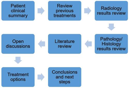
Figure 2 (A) MRI images taken 04 Jan 2020 showing mass near right acetabulum. (B) MRI images take 01 Jun 2020 showing a heterogeneously enhancing mass arising from the right acetabulum, extending into the right hip joint.
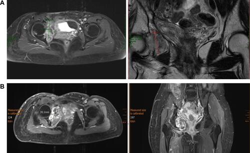
Figure 3 (A) Recurrent pelvic mass with fish-flesh nodules in myxoid lipomatous background. (B) Pelvic mass spindle cell sarcoma. (C) Low-grade sarcoma with pericellular fat infiltration growth pattern typical of DFSP.
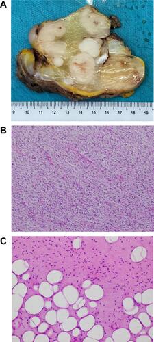
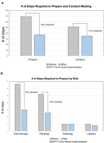
Figure 4 (A) Workflow assessment on the total # of steps assessed before and after implementation of the NAVIFY TB tool. During preparation, the # of steps went from 69 to 37 (46% reduction). For conducting the meeting, the # steps went from 51 to 35 (31% reduction). (B) Preparation step changes before and after implementation of NAVIFY TB. Data manager (33 to 15 steps), pathologist (26 to 13 steps), radiologist (no change), logistics (5 to 4).