
Figures & data

Figure 1 Excision and grafting of the axillary area, with a favorable evolution after Hurley stage III suppurative hidradenitis.
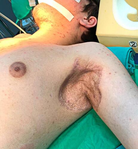
Figure 2 Hurley stage III suppurative hidradenitis, affecting the groin, scrotal, perineal, inner thighs.
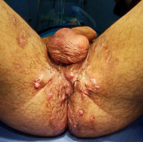
Figure 3 Favorable evolutionary fasciocutaneous flaps under VAC treatment and protection of the perineal area by colostomy. The post-fasciotomy scar is also observed on the right leg.
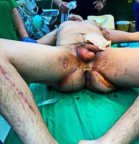
Figure 4 Hyperplasia of the epidermis and the presence of a fistulous tract lined with granulation tissue and hemorrhage (HE stain, x40).
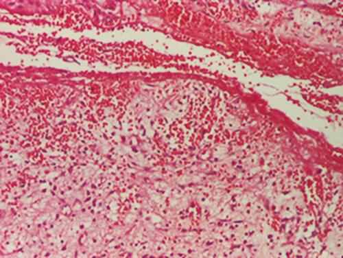
Figure 5 Polymorphic inflammatory infiltrate with polymorphonuclear, lymphocytes, plasmocytes and multinucleous extracellular giant cells (HE stain, x200).
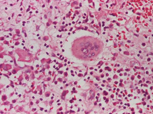
Figure 6 Hemato-fibrino-leukocyte exudate on the surface and fibrosis on the wall (HE stain, x40).
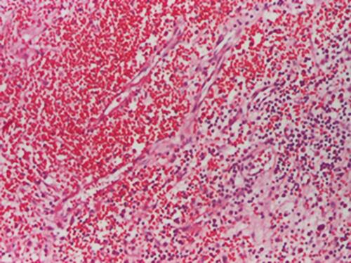
Figure 7 Bilateral axillary hidradenitis, Hurley stage II.

Figure 8 Suppurative hidradenitis in the groin and perineal.
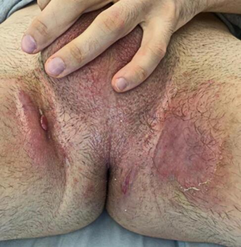
Figure 9 Psoriasis lesions on the elbows and back.
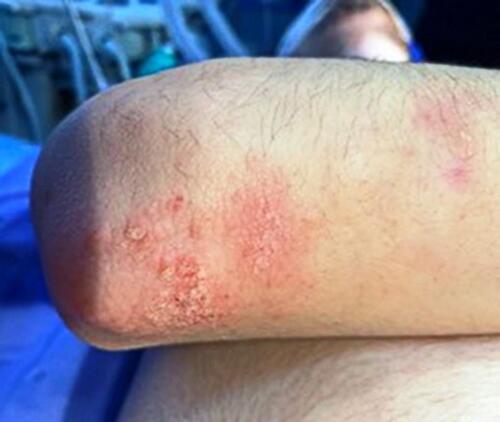
Figure 10 Dextroconvex dorsolumbar scoliosis and L5 spondylolysis.
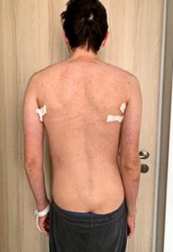
Figure 11 Thickened epidermis, edema, hemorrhagic exudate, inflammatory polymorphic infiltrate (HE stain, x40).
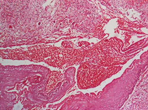
Figure 12 Areas with inflammatory polymorphic infiltrate, with relatively numerous eosinophils throughout the dermis (HE stain, x100).
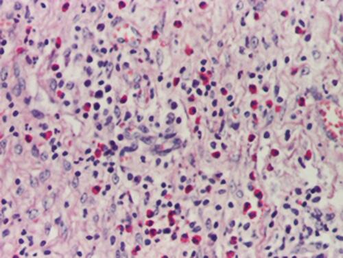
Table 1 Hurley Staging of Suppurative Hidradenitis
Table 2 A Synthesis of the Latest Guidelines for Treatment of HS