Figures & data
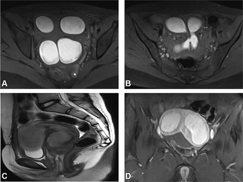
Figure 1 Case of complete septate uterus with urogenital sinus anomaly presenting with cyclic hematuria, categorized by AFS and ESHRE systems. (A) Coronal T2W image demonstrating duplicated cervices. (B and C) Axial T2W images showing complete septate uterus and the narrow connection (arrow) between the vagina and the urinary bladder. (D) Sagittal T2W image showing the dilated proximal vagina with non-visualization of its distal portion. A Gartner duct cyst is seen at its posterior wall.
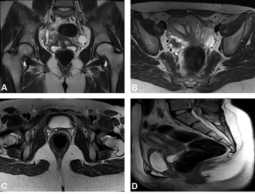
Figure 2 Case of complete septate uterus and vagina down to introitus, categorized by ASRM and ESHRE systems. (A and B) Axial T2W images showing the complete septate uterus and vagina. (C) Coronal T2W image showing the septate cervix and the two vaginae.
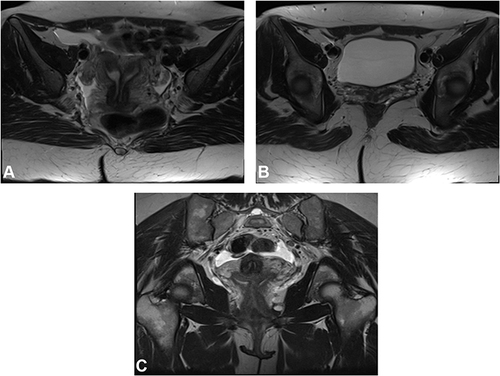
Figure 3 Case of bicornuate bicollis with distal cervical obstruction, categorized only by ESHRE system. (A and B) Axial T1 fat sat images demonstrating marked hematometra and hemoperitoneum (*) with communication between both cervical canals. (C) Sagittal T2W image showing hematometra of the uterine body and cervix. Note the normal vagina. (D) Coronal contrasted T1 fat sat image demonstrating the deep fundal cleft.