Figures & data
Table 1 Comparison of the Baseline Data Between the Three Groups
Table 2 Comparison of the Baseline Data Between the Two Groups
Figure 1 Bedside ultrasound. (A-H): Right pleural effusion is the accumulation of fluid in the area between the layers of the pleura around the right lung, pleural effusion is the separation of the walls of the pleural cavity, and the space is filled with fluid, hydropneumothorax is the unusual accumulation of air and fluid in the pleural space and pneumothorax shows significant lung patches, decreased lung movement, and stratosphere symptoms.
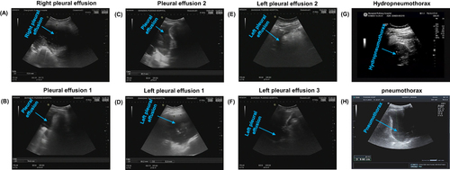
Table 3 Time to Diagnose and Treat in the Three Patients’ Group
Table 4 Patients’ Diagnostic Rate in Each Examination Method Group
Table 5 LUS Scores and Diaphragmatic Displacement are Compared Between the Two Groups
Table 6 Lung Ultrasonography Prognostic LUS Score and Diaphragm Displacement in Patients with Severe Multiple Injuries Complicated by Respiratory Failure
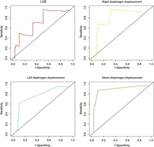
Figure 2 ROC curve showing prediction results for various markers.