
Figures & data
Table 1 Acquisition Parameters and Scan Times for Conventional and Ultrafast Protocols
Figure 1 The example of sagittal T1-weighted (T1WI) and T2-weighted (T2WI), and axial T2-weighted images, obtained from conventional, ultrafast and ultrafast-DL-3DIIE protocols.
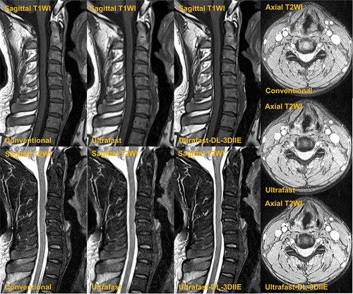
Table 2 Qualitative Assessment and Comparison
Table 3 Quantitative Assessment and Comparison
Table 4 Integrated Results of Diagnostic Performance by Two Observers
Figure 2 Sagittal T2-weighted images of a participant with central canal stenosis (C3-7 levels, Orange ellipses), were obtained from both conventional (a) and ultrafast-DL-3DIIE (b) protocols. And sagittal T2-weighted images of a participant with foraminal stenosis (C5-6 level, Orange arrows), obtained from both conventional (c) and ultrafast-DL-3DIIE (d) protocols.
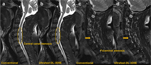
Figure 3 Sagittal T2-weighted images of a participant with disc degeneration (C3-4, C4-5 and C5-6 discs, Orange ellipses), were obtained from both conventional (a) and ultrafast-DL-3DIIE (b) protocols. And sagittal T2-weighted images of a participant with disc herniation (C5-6 disc, Orange arrows), obtained from both conventional (c) and ultrafast-DL-3DIIE (d) protocols.
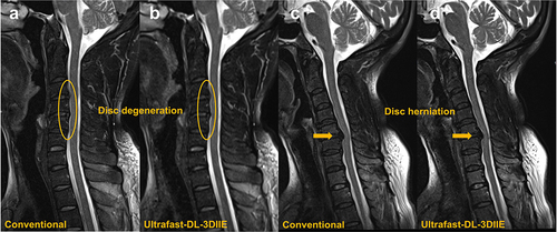
Table 5 Interchangeability Results for Two Protocols