
Figures & data
Figure 1 Multidisciplinary approach to MD.
Abbreviations: GI, gastrointestinal; MD, Menkes disease.
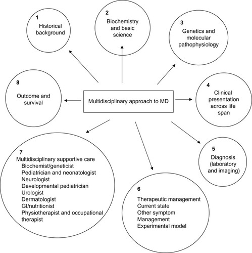
Figure 2 MD: (A) scalp shows “kinky hair”, (B) the inset shows “pili torti” and a normal hair strand under a high power microscope.
Abbreviation: MD, Menkes disease.
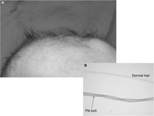
Table 1 Copper enzymes and their role in MD
Table 2 Functions of ATP7A: a multitasking protein in the nervous system
Table 3 Manifestations of MD
Table 4 Evolution and progression of epilepsy in MD
Figure 3 EEG segments from patient with MD.
Notes: (A) Anterior–posterior bipolar montage shows ictal rhythm corresponding to a focal seizure originating in the right mid to posterior temporal regions (15 mm/s, sensitivity at 15 μV/mm). (B) Ictal rhythms showing rhythmic generalized spike and slow waves (time base at 30 mm/s, sensitivity at 20 lV/mm). (C) Late stages – interictal background rhythms are slow for age, with multifocal independent spikes and spike and wave complexes (time base at 30 mm/s, sensitivity at 20 lV/mm).
Abbreviations: EEG, electroencephalography; MD, Menkes disease.
Abbreviations: EEG, electroencephalography; MD, Menkes disease.
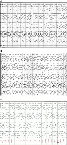
Figure 4 MRI of the brain (A) and MRA (B) from the same patient.
Notes: (A) Generalized fronto-temporal cerebral atrophy resulting in exposure of the insula, and thinning of the subcortical white matter (arrows). (B) Tortuosity of intracranial vertebral arteries (arrow).
Abbreviations: L, left; MRA, magnetic resonance angiography; MRI, magnetic resonance imaging; R, right.
Abbreviations: L, left; MRA, magnetic resonance angiography; MRI, magnetic resonance imaging; R, right.
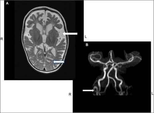
Table 5 Multidisciplinary management in MD