Figures & data
Table 1 Diagnostic criteria sets for BS, ISG and ICBD
Figure 1 Mucocutaneous lesions of Behçet’s syndrome.
Notes: (A) OU; (B) GU with a scar; (C) PPL; (D) acne with arthritis; (E) EN and (F) leg ulcers.
Abbreviations: OU, oral ulcer; GU, genital ulcer; PPL, papulopustular lesions; EN, erythema nodosum.
Abbreviations: OU, oral ulcer; GU, genital ulcer; PPL, papulopustular lesions; EN, erythema nodosum.
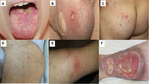
Figure 2 (A, B) Axial FLAIR-T2 images reveal hyperintense lesion in the brainstem (arrow), (C) axial T1 gadolinium sequence shows gadolinium enhancement (arrow) and (D) prominent brainstem atrophy (arrow) on the follow-up MRI after 3 years.
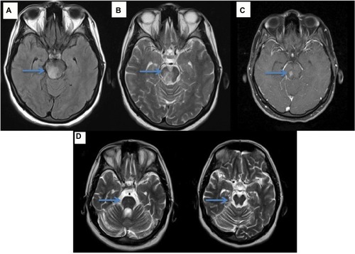
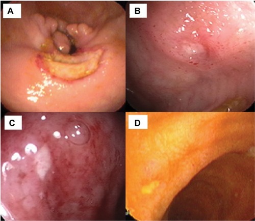
Figure 3 Endoscopic appearance of round ulcers of GI involvement of BS.
Notes: (A, B) Some of these ulcers can be deep with elevated edges and (C, D) some are shallow.
Abbreviations: GI, gastrointestinal involvement; BS, Behçet’s syndrome.
Abbreviations: GI, gastrointestinal involvement; BS, Behçet’s syndrome.