Figures & data
Table 1 Patient characteristics
Table 2 The assessment of pain relief during treatment period
Table 3 The assessment of immediate and long-term pain relief after treatment
Figure 1 Detection of the neuromas.
Notes: (A) Using ultrasound probe to scan the stump limb. (B) The transverse axial view of neuroma. (C) The longitudinal axial view of neuroma. The dotted line indicates neuroma.
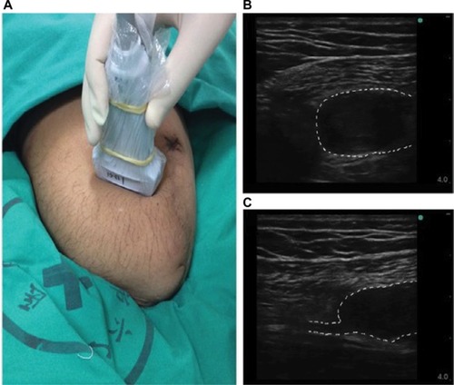
Figure 2 Representative images of alcohol neurolysis to the neuromas.
Notes: The injection needle was inserted into the neuroma body, and the tip was adjusted to evoke the extreme pain. The dotted line indicates neuroma.
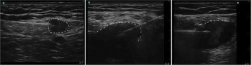
Figure 3 RF procedure.
Notes: (A) The RF generator machine. (B) The transverse axial view of neuroma body. (C) RF needle was advanced to the responsible nerve 5 mm away from neuroma stalk. The dotted line indicates neuroma.
Abbreviation: RF, radiofrequency.
Abbreviation: RF, radiofrequency.
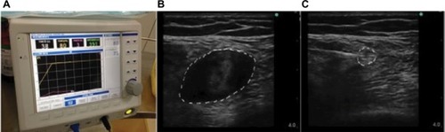
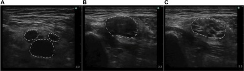
Figure 4 The variation of neuromas.
Notes: (A) Three neuromas origin from 1 nerve. (B and C) The nerve immediately proximal to the neuroma is pathological. The nerve is usually swollen, with its diameter as large as 1 cm or more. (B) is the transverse axial view of neuroma body, and (C) is the transverse axial view of neuroma stalk. The dotted line indicates neuroma.