Figures & data
Figure 1 Genotyping and autotomy evaluation after axotomy.
Notes: (A) Identification of KO (−/−), heterozygous (+/−) and WT (+/+) mice by PCR. (B) Onset of autotomy observed on days 0, 1, 3 and 7 post-surgery in KO and WT mice. (C) Percentage of autotomized mice on day 7 post-surgery. (D) Representation of the severity of autotomy by score in KO and WT mice on day 7 post-surgery. ***p < 0.001.
Abbreviations: KO, knockout; PCR, polymerase chain reaction; WT, wild-type.
Abbreviations: KO, knockout; PCR, polymerase chain reaction; WT, wild-type.
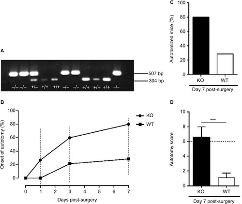
Figure 2 Autotomy detected 7 days after axotomy.
Notes: (A, B) Schematics of autotomy occurred on the hindpaws of WT (A) and KO (B) mice. The numbers 1–8 represent different individuals in the experiments. Black and green arrows indicate the injury of nails and toes, respectively, and blue arrows indicate bleeding spots.
Abbreviations: KO, knockout; WT, wild-type.
Abbreviations: KO, knockout; WT, wild-type.
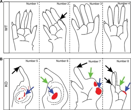
Figure 3 Neuronal number of L5 DRGs 1 month after axotomy.
Notes: (A) The number of neurons is significantly decreased in the ipsilateral DRGs as compared with the contralateral DRGs in both KO (n = 7) and WT (n = 7) mice, respectively. There are significantly more neurons in the contralateral DRGs in KO mice than in WT mice. The number of neurons in the ipsilateral DRGs of KO mice is similar to that of WT mice. *p < 0.05; ***p < 0.001; ns p > 0.05. (B) Immunofluorescence micrograph presents double labeling with CGRP anti-serum (green) and PI (red) in intact DRG in WT mouse. PI-labeled neurons (arrows) and satellite cells (arrowheads) exhibit a strong and typical cytoplasmic and nuclear fluorescence, whereas CGRP-LI is exclusively present in the cytoplasm and fibers (green). CGRP-LI is detected in small (I), medium (II) and large (III) NPs. (C) Immunofluorescence micrograph presents TUNEL-positive cells in axotomized DRG in WT mouse. Thick arrow points to a TUNEL-positive neuron, thin arrows point to TUNEL-positive satellite cells and a double arrowhead points to a TUNEL-negative neuron. Scale bars, B = 5 μm and C = 10 μm.
Abbreviations: CGRP, calcitonin gene-related peptide; DRG, dorsal root ganglion; KO, knockout; L5, lumbar-5; NPs, neuron profiles; ns, no significance; PI, propidium iodide; TUNEL, terminal deoxynucleotidyl transferase-mediated dUTP nick-end labeling; WT, wild-type.
Abbreviations: CGRP, calcitonin gene-related peptide; DRG, dorsal root ganglion; KO, knockout; L5, lumbar-5; NPs, neuron profiles; ns, no significance; PI, propidium iodide; TUNEL, terminal deoxynucleotidyl transferase-mediated dUTP nick-end labeling; WT, wild-type.
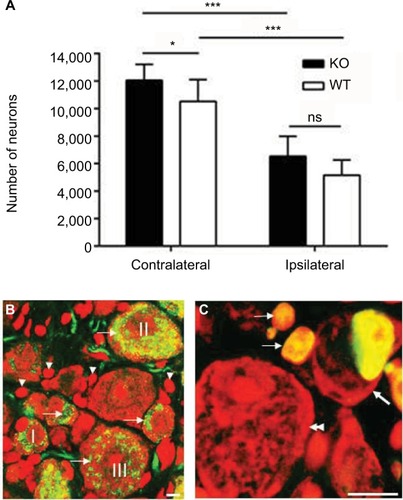
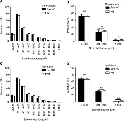
Figure 4 Soma size distribution of DRG NPs 1 month after axotomy.
Notes: Size distribution of neuron profiles is not different between KO and WT mice in the contralateral (A, B) or the ipsilateral (C, D) DRGs, respectively. ns (p > 0.05). n=200 NPs per group.
Abbreviations: DRG, dorsal root ganglion; KO, knockout; NPs, neuron profiles; ns, no significance; WT, wild-type.
Abbreviations: DRG, dorsal root ganglion; KO, knockout; NPs, neuron profiles; ns, no significance; WT, wild-type.