Figures & data
Figure 1 Flowchart showing overview of the protocols for the in vivo ovine studies.
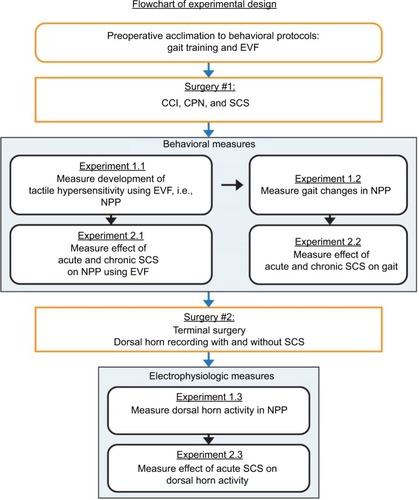
Figure 2 (A) Dissection and isolation of the peroneal nerve at the level of the stifle joint. The nerve is palpable just behind the stifle joint, coursing around the fibular head. The stifle and hock joints are marked for reference, in addition to the peroneal nerve. (B) Four suture ligatures have been placed around the nerve to create a 25% constriction (i.e., a reduction in the diameter of the constricted nerve from the normal diameter by 25%).
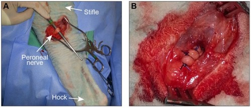
Figure 3 (A) Relative to a mock spinal column, from left to right are shown the micrometer-driven translation stage used to control the microelectrode insertion depth and position, the mounting tower, and the Teflon® threaded plug used to seal the tower interior when not in use. (B) Top–down view of the apparatus from (A) anchored in place on the sheep spinal column for a dorsal horn recording. (C) H&E stain of an axial cross section of the dorsal portion of the spinal cord at the level of the lumbar enlargement (L6). The white matter tracts of the spinal cord are slightly more eosinophilic, and the neuron-containing gray matter shows the characteristic butterfly wing structure. The dashed rectangle outlines the targeted recording region within the dorsal horn. The DF, DH, LF, and the central canal are marked. (D) Example recording of a single neuron’s activity recorded at a depth of 1414 µm.

Figure 4 Withdrawal thresholds of the hind limbs in three sheep as measured with von Frey filaments and anesthesiometer from (A, B) one animal with a sectioned nerve and (C, D) two animals with constriction nerve injuries.
Abbreviation: SCS, spinal cord stimulation.
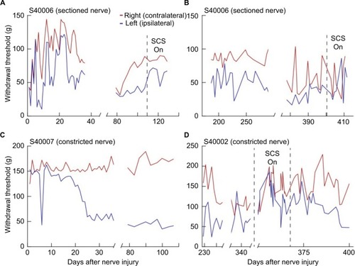
Figure 5 The results for the von Frey measurements for the five sheep included in the final protocol (#40014, #40015, #40016, #40021, and #40022) are presented in and expressed in terms of means and standard errors.
Abbreviation: SCS, spinal cord stimulation.
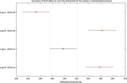
Table 1 von Frey thresholds of five sheep in standardized protocol
Figure 6 (A) Range of flexion–extension angle of the hock joint in the sagittal plane (leg swing angle). (B) Angular speed during the first 100 ms of the gait cycle appears to increase in the contralateral (right) limb for the sheep with the cut peroneal nerve (#40006), but this does not occur for the sheep with the constricted nerves (#40002 and #40007).
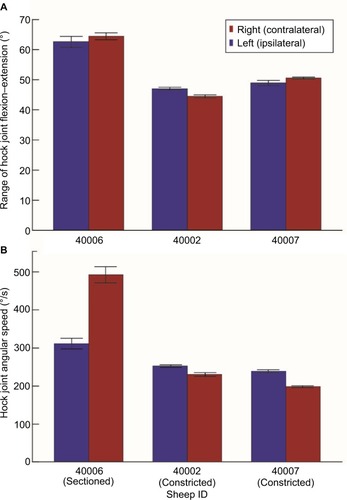
Figure 7 Mean height of both back hooves plotted against percent of gait cycle for the sheep with a cut peroneal nerve (#40006).
Abbreviation: SCS, spinal cord stimulation.
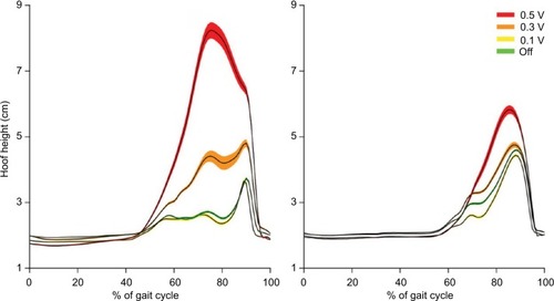
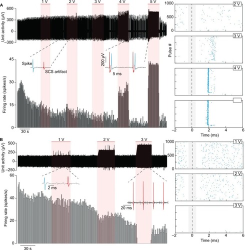
Figure 8 Example recordings of dorsal horn neurons (A) without spontaneous activity and (B) with spontaneous activity.
Abbreviation: CCI, chronic constriction injury.
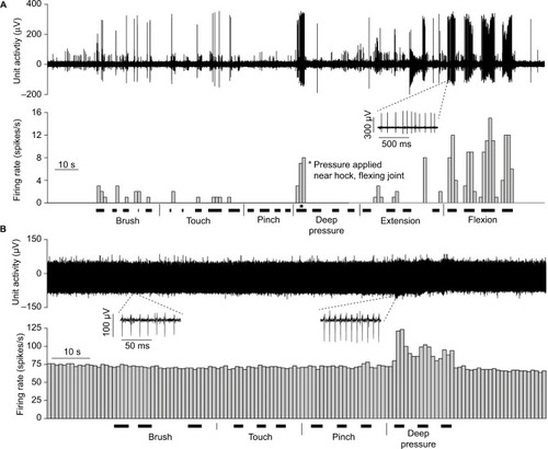
Figure 9 Neuronal activity of two neurons from different blocks recorded during SCS from the same sheep (#40022).
Abbreviation: SCS, spinal cord stimulation.