
Figures & data
Figure 1 Photograph and the corresponding illustration showing five branches of the SCN on the left side of a specimen obtained from the cadaver of a 90-year-old woman (specimen no. 8). The most lateral branch (a5) had separate origins from the T12 and L2 nerve roots. The third most medial branch (a3) had separate origins from the L2 and L3 nerve roots.
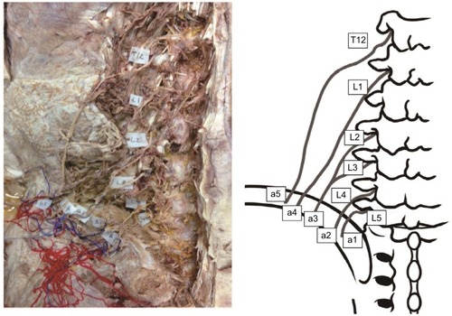
Figure 2 Photographs and the corresponding illustration showing three branches of the SCN on the right side of a cadaveric specimen obtained from an 89-year-old man (specimen no. 22). The L5 nerve roots had a dorsal branch ramifying into the most medial SCN branch (a1) and the second most medial branch (a2). The lateral branch of the SCN (a3) had separate origins from the L2 and L3 nerve roots.
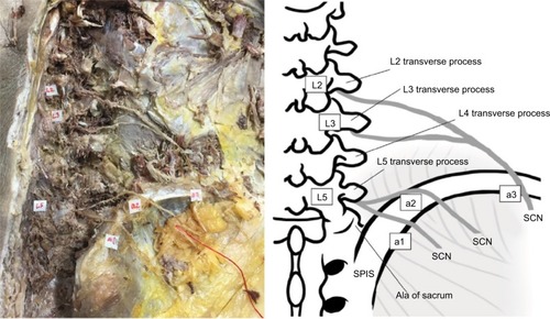
Table 1 Levels of lumbar nerve root originating SCN branches
Figure 3 Photographs showing entrapped branches of the medial branch of the SCN in cadaveric specimen no. 23. a2 is the second most medial branch of SCN.
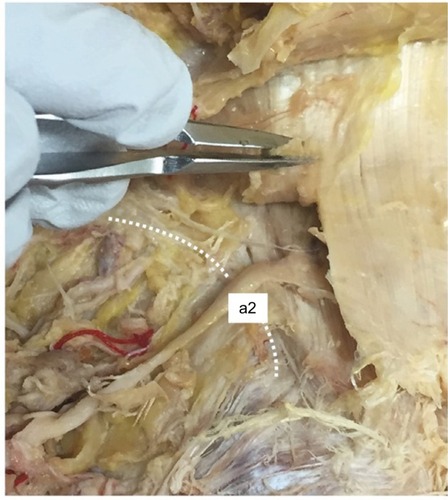
Table 2 Distances from anatomical landmarks and diameter of superior cluneal nerves