
Figures & data
Figure 1 Body manikins used to define the 45 pain sites in the front and the back
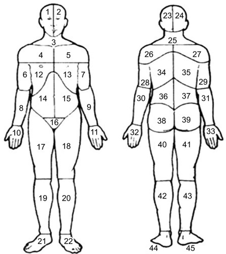
Figure 2 Preshaded manikins used to define the 23 APSs; each area of the same color corresponds to the same APS in the front and the back of the body.
Abbreviation: APSs, anatomical pain sites.
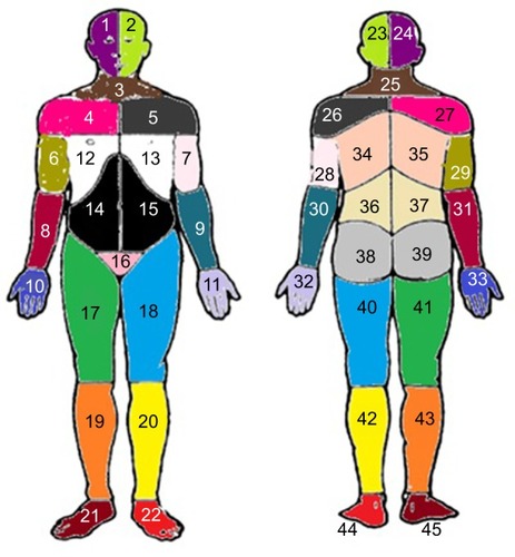
Table 1 Distribution of sociodemographic characteristics and all studied variables in the whole sample (n = 6,611)Table Footnotea
Figure 3 Distributions of the number of APSs categorized into 0, 1, 2, to 23 pain sites in males, females, and the total sample.
Abbreviation: APSs, anatomical pain sites.
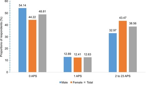
Figure 4 Distributions of the location of the APSs in males and females.
Abbreviation: APSs, anatomical pain sites.
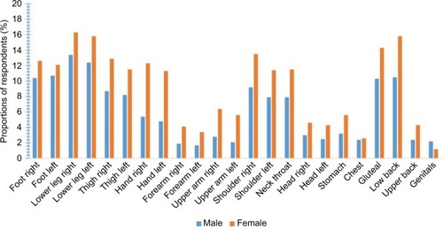
Table 2 The total number of APSs and the location of the APSs in the whole sample
Table 3 Results of regression analyses: cross-sectional associations of the factors associated with the number of APSs
Table S1 Results of regression analyses: cross-sectional associations of the factors associated with the NPS (0–45)