Figures & data
Figure 1 (A) Schematic diagram showing the probe position and needle direction for greater occipital nerve block. (B) Ultrasound image demonstrating needle placement and dye spread of GON block.
Abbreviations: GON, greater occipital nerve; SP, spinous process; OCI, obliquus capitis inferior; TON, third occipital nerve.
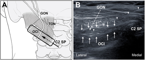
Table 1 Comparison of dye staining between ultrasound-guided GON block with 1 mL and 5 mL of dye solution
Figure 2 Comparison of the dye-spreading patterns between GON block using 1 mL (left) and 5 mL (right) of dye solution.
Abbreviations: GON, greater occipital nerve; TON, third occipital nerve; OCI, obliquus capitis inferior.
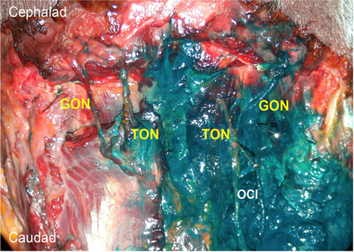
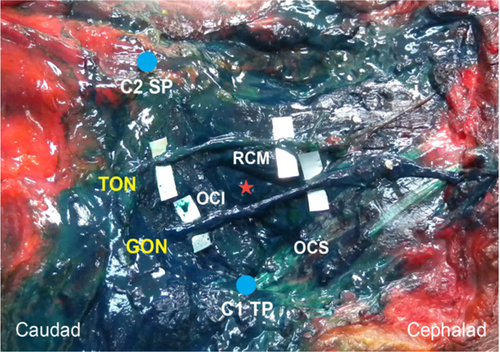
Figure 3 Suboccipital region after GON block using 5 mL of dye solution.
Note: The red asterisk indicates the suboccipital triangle.
Abbreviations: GON, greater occipital nerve; TON, third occipital nerve; OCI, obliquus capitis inferior; OCS, obliquus capitis superior; RCM, rectus capitis posterior major; SP, spinous process; TP, transverse process.
Abbreviations: GON, greater occipital nerve; TON, third occipital nerve; OCI, obliquus capitis inferior; OCS, obliquus capitis superior; RCM, rectus capitis posterior major; SP, spinous process; TP, transverse process.