
Figures & data
Figure 1 US-guided injections with demonstration of needle tip (long arrow) along with median nerve (short arrow) just before the carpal tunnel inlet. a) Ulnar in-plane approach: in this method, the wrist was placed on the table in slightly dorsiflexed and supinated position and the injection was done at tunnel inlet from ulnar side lateral to ulnar artery, below the median nerve. b) Midline in-plane approach: in this method, we placed the wrist on the same position as above mentioned and the same needle features but the injection was done above the median nerve.
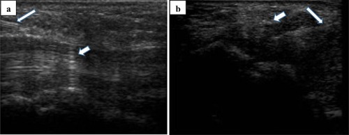
Figure 2 US-measuring of median nerve (white arrow) just before the carpal tunnel inlet.
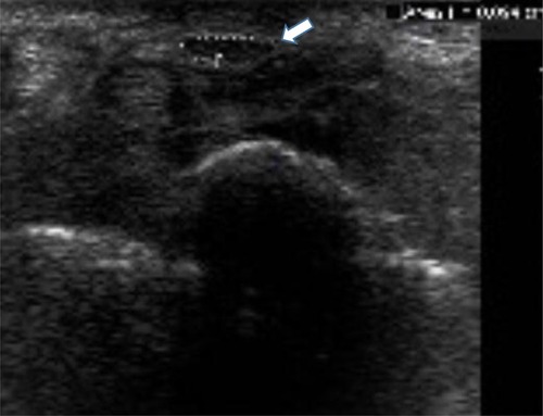
Figure 3 Flowchart of the study’s population.

Table 1 Comparison Of Demographic Characteristics And Baseline Variables Among Patients Of Three Groups
Table 2 Comparison Of Pre- And Post-Treatment Clinical Values In Each Group
Table 3 Comparison Of Pre- And Post-Treatment Electro-Diagnostic Values In Each Group
Table 4 Comparison Of Improvement In Clinical And Electro-Diagnostic Variables Among Three Groups