Figures & data
Figure 1 Doppler ultrasonography of left knee posterior side, the dotted circle is P2 segment occlusion with thrombosis formation at the popliteal artery.
Abbreviations: PA, popliteal artery; PV, popliteal vein; MHG, medial head of gastrocnemius muscle.
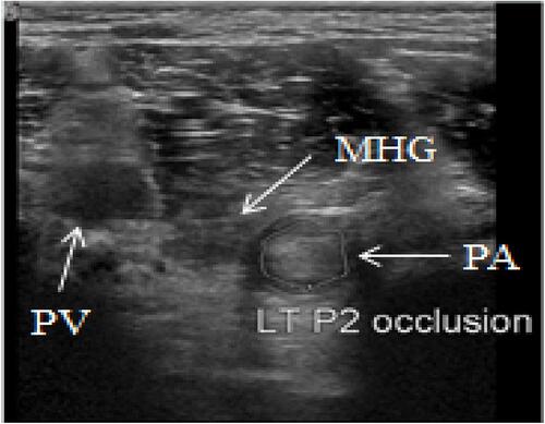
Figure 2 (A) The left knee coronal T2 with contrast MRI, (B) the popliteal artery compressed by the medial head of the gastrocnemius muscle to thicken the vessel wall.
Abbreviations: PA, popliteal artery; PV, popliteal vein; MHG, medial head of gastrocnemius muscle.
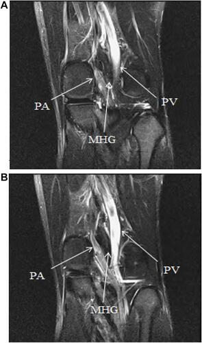
Figure 3 The left knee sagittal T1 with contrast MRI; aneurysm formation caused by the compressed popliteal artery can be observed at the popliteal artery.
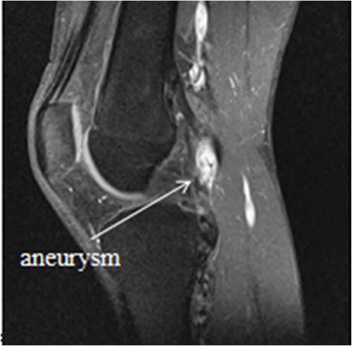
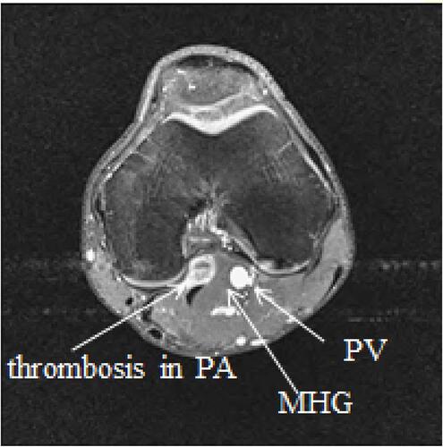
Figure 4 The left knee axial T1 with contrast MRI; thrombosis formation in the vessel wall of the popliteal artery can be observed.
Abbreviations: PA, popliteal artery; PV, popliteal vein; MHG, medial head of gastrocnemius muscle.