Figures & data
Table 1 Demographic Data and Clinical Characteristics of the LBLP and HCs Groups
Table 2 Differences in the Alterations of sFC of the S1 Cortex Between the LBLP and HCs Groups (Two-Sample t-Test, Two-Tailed, Voxel-Level P < 0.001, GRF Correction, Cluster-Level P < 0.005)
Figure 1 Static functional connectivity spatial distributions were observed at the group level for LBLP patients and healthy controls (the left somatotopic S1 subregions).
Abbreviations: sFC, static functional connectivity; S1, primary somatosensory cortex; LBLP, low-back-related leg pain; HCs, healthy controls.
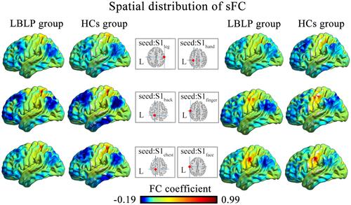
Figure 2 Differences in the alterations of sFC between the LBLP patients and healthy controls (two-sample t-test, two-tailed, voxel-level P < 0.001, GRF correction, cluster-level P < 0.005).
Notes: (A–C) Patients with LBLP exhibited increased sFC to the right SFG/MFG. (D) Patients with LBLP exhibited increased sFC to the right MFG. Values of sFC are the mean ± SEM.
Abbreviations: sFC, static functional connectivity; LBLP, low-back-related leg pain; HCs, healthy controls; SFG, superior frontal gyrus; MFG, middle frontal gyrus; S1back_L, representation of the left back in the primary somatosensory cortex.
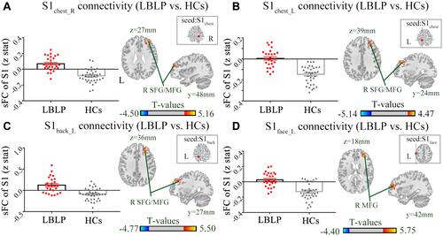
Table 3 Differences in the Alterations of dFC of the S1 Cortex Between the LBLP and HCs Groups (Two-Sample t-Test, Two-Tailed, Voxel-Level P < 0.01, GRF Correction, Cluster-Level P < 0.05)
Figure 3 Differences in the alterations of dFC between LBLP patients and healthy controls (two-sample t-test, two-tailed, voxel-level P < 0.01, GRF correction, cluster-level P < 0.05).
Notes: (A) Increased dFC between the right S1hand cortex and the left PRG/POG in LBLP patients. (B) Increased dFC between the right S1finger cortex and the right PRG/POG in LBLP patients.
Abbreviations: dFC, dynamic functional connectivity; LBLP, low-back-related leg pain; HCs, healthy controls; PRG, precentral gyrus; POG, postcentral gyrus; S1hand_R, representation of the right hand in the primary somatosensory cortex; values of sFC are the mean ± SEM.
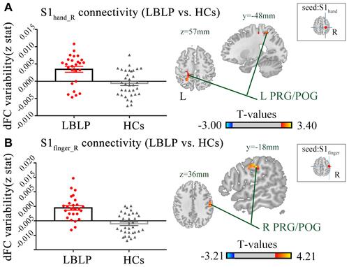
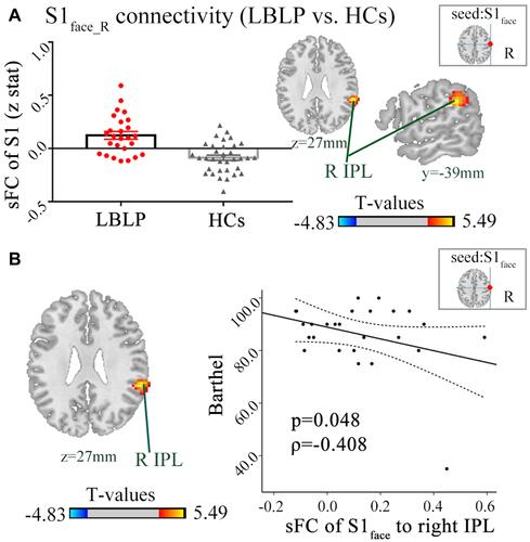
Figure 4 (A) Patients with LBLP exhibited increased sFC to the right IPL. Values are the mean ± SEM (two-sample t-test, two-tailed, voxel-level P < 0.001, GRF correction, cluster-level P < 0.005). (B) Partial correlational analysis between sFC alterations and the Barthel index in LBLP patients.
Abbreviations: sFC, static functional connectivity; LBLP, low-back-related leg pain; IPL, inferior parietal lobule; S1face_R, representation of the right face in the primary somatosensory cortex.