
Figures & data
Figure 1 PRF procedures. (A) Location for puncture. (B–F) Consecutive CT scans showing the puncture needle gradually reaching the medial edge of styloid process. Patients were placed in a supine position with head slightly turned contralaterally. White arrows indicate the needle, white triangles indicate the styloid process.
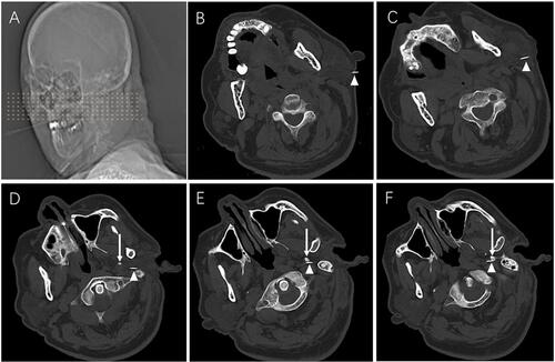
Table 1 Likert Scale
Table 2 The Landriel Ibanez Classification of Complications
Table 3 The Demographic Data and Baseline Characteristics of GPN Patients
Figure 2 Kaplan–Meier recurrence-free survival curves for idiopathic glossopharyngeal neuralgia patients who underwent CT-guided pulsed radiofrequency.
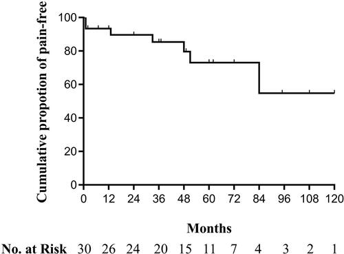
Figure 3 Flow Chart.
Abbreviations: GPN, glossopharyngeal neuralgia; PRF, pulsed radiofrequency; MVD, microvascular decompression.
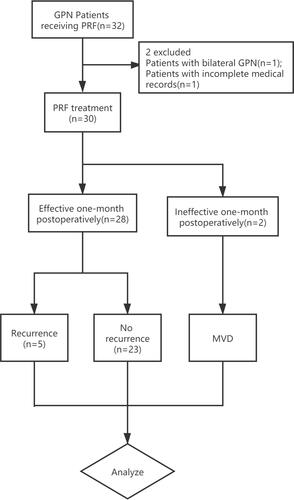
Table 4 Complications of PRF for 30 Cases