Figures & data
Figure 1 Examples of APs recorded from mechanoreceptive neurons. (A) Representative intracellular somatic action potential of an A-fiber neuron evoked by electrical stimulation of the dorsal root showing the electrophysiological parameters measured, including: 1, resting membrane potential; 2, action potential duration at base; 3, action potential rise time; 4, action potential fall time; 5, action potential amplitude; 6, AHP duration to 50% recovery; 7, and afterhyperpolarization amplitude below Vm. In addition, maximum rising and falling rates, (dV/dt) max, were measured from the differential trace of the action potential. (B) Somatic action potentials evoked by dorsal root stimulation and recorded intracellularly from 12 mechanoreceptive neurons selected to represent the mean action potential duration values for each of the different groups of neurons in control (upper) and neuropathic (lower) animals. The action potential duration and conduction velocity for each neuron are given below each record. The horizontal lines across the action potentials indicate zero membrane potential.
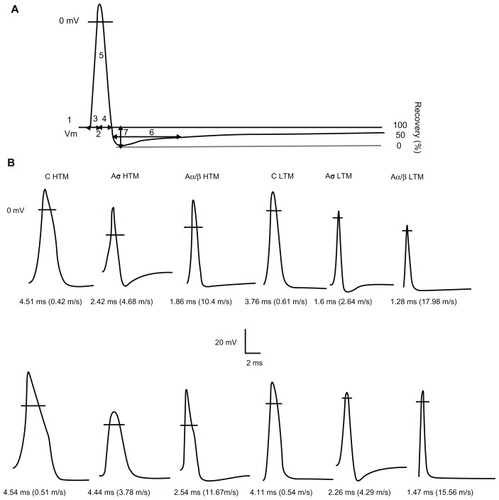
Figure 2 Comparison of action potential resting membrane potential and amplitude of dorsal root ganglion neurons between control and neuropathic rats. Scatter plots show the distribution of the variables with the median (horizontal line) superimposed in each case.
Abbreviations: HTM, high threshold mechanoreceptor neurons; LTM, low threshold mechanoreceptor neurons; UN, unresponsive neurons; CUT, Aβ LTM including guard/field hair neurons, rapidly adapting neurons and slowly adapting neurons; MS, Aβ LTM muscle spindle neurons.
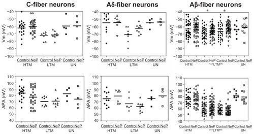
Figure 3 Comparison of action potential dynamic parameters of dorsal root ganglion neurons between control and neuropathic rats. Scatter plots show the distribution of the variables with the median (horizontal line) superimposed in each case.
Abbreviations: APdB, action potential duration at base; APRT, action potential rise time; APFT, action potential fall time; MRR, maximum AP rising rate; MFR, maximum AP falling rate; HTM, high threshold mechanoreceptor neurons; LTM, low threshold mechanoreceptor neurons; UN, unresponsive neurons.
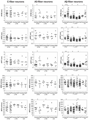
Figure 4 Comparison of AHP variables of DRG neurons between control and neuropathic rats. Scatter plots show the distribution of the variables with the median (horizontal line) superimposed in each case.
Abbreviations: AHPA, afterhyperpolarization amplitude; AHP50, afterhyperpolarization duration to 50% recovery; HTM, high threshold mechanoreceptor neurons; LTM, low threshold mechanoreceptor neurons; UN, unresponsive neurons.
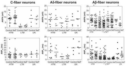
Table 1 Comparison of properties of high-threshold mechanoreceptive and low-threshold mechanoreceptive dorsal root ganglion neurons between control and neuropathic rats
Figure 5 Comparison of dorsal root conduction velocity of dorsal root ganglion neurons between control and neuropathic rats. Scatter plots show the distribution of variables with the median (horizontal line) superimposed in each case. Details are the same as in .
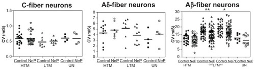
Table 2 Locations of receptive fields of high-threshold mechanoreceptors and low-threshold mechanoreceptors in controls and neuropathic rats