Figures & data
Figure 1 Dimensions of the masseter muscle in relation to segments: (A) superior transverse; (B) lower transverse; (C) longitudinal line of the masseter muscle; and (D) middle transverse line.
Note: Figure courtesy of Marcos Vinicius de Oliveira.
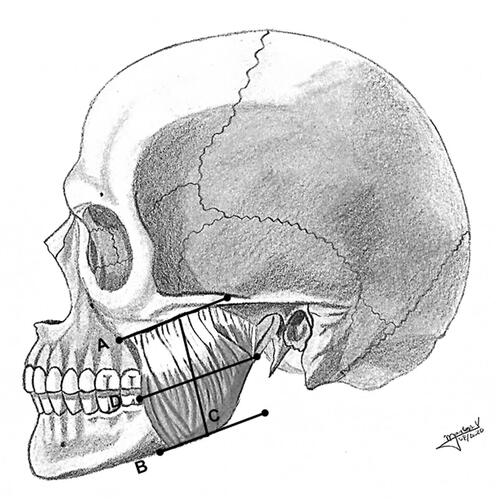
Figure 2 (A) Left side view of the face. Orientation of the abscissa (x) and ordinate (y) of the left masseter. By convention: upper anterior quadrant (UAQ) with positive ordinate and abscissa; lower posterior quadrant (LPQ) with negative ordinate and abscissa; superior posterior quadrant (SPQ) with negative and positive ordinate abscissa; and lower anterior quadrant (LAQ) with positive and negative ordinate abscissa; (B) Left side view of the face. Sextant of the left masseter muscle for localization of the penetration points of the masseteric nerve. D: The middle transverse line separates the upper and lower areas divided into three equally sized segments. The areas are numbered from I–VI, where I–III are superior and IV–VI are inferior in the posterior to anterior direction, respectively; (C) Left side view of the face. 1) zygomatic bone; 2) jaw; 3) temporal muscle; 4) folded masseter muscle. A) Masseteric nerve indicated in yellow; B, C, D and E indicate the branches of the masseteric nerve entering the masseter muscle in white.
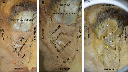
Table 1 Description of the Characteristics of Cadavers
Table 2 Description of Masseter Measurements by Sex
Table 3 Description of the Entry Points of the Masseteric Nerve Branches in the Masseter Muscle According to Sex and Side
Table 4 Description of the Number of Points of Entry of the Masseteric Nerve Branches According to Sextants
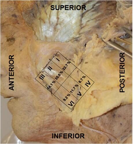
Figure 3 Sextant left masseter muscle. Percentage of nerve entry points by area. Areas numbered from I–VI, where I–III are superior and IV–VI are inferior in the posterior to anterior direction, respectively.