
Figures & data
Figure 1 (A, E and G) CT-guided path design for T1, T2, and T3 puncture; (B) the tip of the puncture needle reached the T1 costal transverse joint; (C) the puncture needle have successfully crossed the costal transverse joint; (D, F and H) CT images of T1, T2, and T3 puncture, finally and respectively; (I) CT three-dimensional reconstruction of T1, T2, T3 puncture.
Notes: The red arrow indicates the current position reached by the puncture needle, and the green arrow indicates the costal transverse process joint.
Abbreviations: T1, the intervertebral foramen of the first thoracic vertebra; T2, the intervertebral foramen of the second thoracic vertebra; T3, the intervertebral foramen of the third thoracic vertebra.
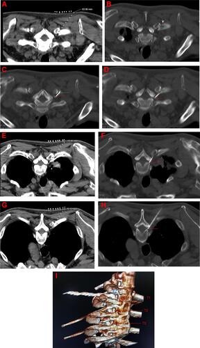
Table 1 Clinical Characteristics of Patients
Table 2 Comparison of Preoperative and Postoperative NRS
Table 3 Comparison of Preoperative and Postoperative Medication