Figures & data
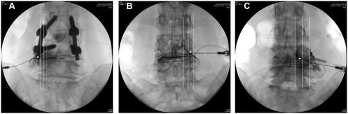
Figure 1 Fluoroscopic images of selective epidural transforaminal epidural injection (STE). (A) skin entry point was just lateral to ipsilateral superior articular process of the lower vertebrae of the target level (asterisk) on oblique view. (B) The needle was inserted with tunnel view technique until it can be orientated. (C) The needle was advanced to the lateral margin of the pedicle in the AP view. (D) The needle was advanced to pass beyond the front half of the vertebral foramen and anterior epidural space was confirmed by contrast media.
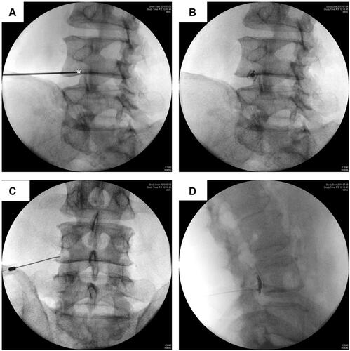
Figure 2 Needle tip position: (A) extraforamen, outside the pedicle; (B) lateral foramen (LF), lateral to the interpedicular line; and (C) medial foramen (MF), medial to the interpedicular line.
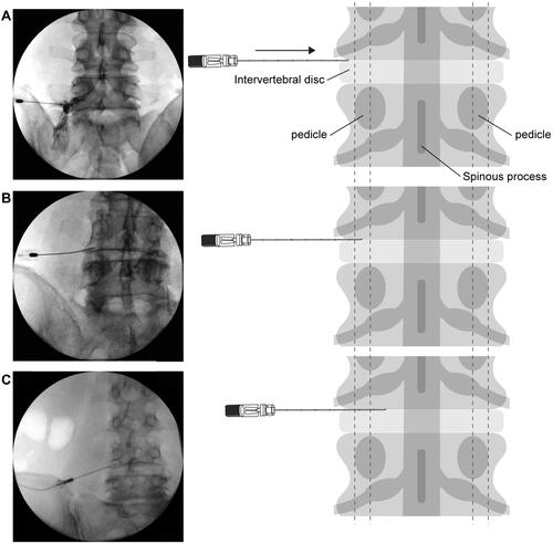
Figure 3 Contrast media pattern. (A) Nerve root: the contrast media is dispersed along the nerve root only. (B) Epidural: contrast media is dispersed into the epidural space.
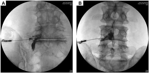
Figure 4 Flowchart of cases. Regarding abnormal patterns, two EF cases show intradiscal pattern; of the three LF cases, two show intradiscal pattern and one shows intravascular pattern; of the three MF cases, two show intradiscal pattern and one shows intrathecal pattern.
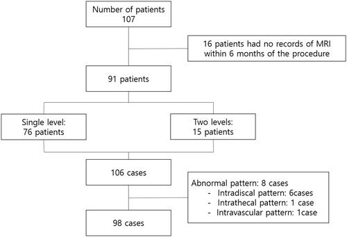
Table 1 Sites of Selective Transforaminal Epidural Injection
Table 2 Lumbar Spinal Fusion Surgery History, Level of Procedure, and MRI Findings According to Tip Position
Table 3 Contrast Media Dispersion Patterns According to Needle Tip Position
Table 4 Logistic Analysis for Factors Associated with Contrast Media Dispersion into the Epidural Space
Table 5 Contrast Media Dispersion Pattern According to Needle Tip Position in Patients with Grade 3 Foraminal Stenosis and Grade 3 Lateral Recess Stenosis (n = 15)
Figure 5 Intradiscal patterns (A) in extraforamen (EF) (B) in lateral foramen (LF) (C) in medial foramen (MF). White dot: position of the needle tip. White lines: medial margin, half line, lateral margin of the pedicle.