Figures & data
Figure 1 Experimental design.
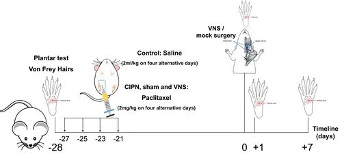
Figure 2 The effects of vagus nerve stimulation (VNS) on heat and mechanical hyperalgesia in a chemotherapy-induced peripheral neuropathy (CIPN) model. CIPN was induced in rats by paclitaxel treatment on four alternative days. Rats of the VNS group underwent VNS on day 0, whereas rats of the sham group underwent mock surgery. Behavioral tests were performed on days +1 and +7 of the experiment. (A) Hindpaw withdrawal latencies during the plantar test. (B) Hindpaw withdrawal frequencies during the 15-g von Frey hair test. n = 12 per group per time point for both panels. *p<0.05 and **p<0.001.
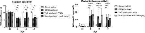
Figure 3 Effect of vagus nerve stimulation (VNS) on levels of pro- and anti-inflammatory factors. Protein was extracted from dorsal root ganglia of CIPN rats treated with VNS or not. (A, B) Representative Western blot and corresponding densitometry on day +1. (C, D) Representative Western blot and corresponding densitometry on day +7. n = 7 per group per time point. * p<0.05.
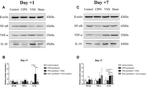
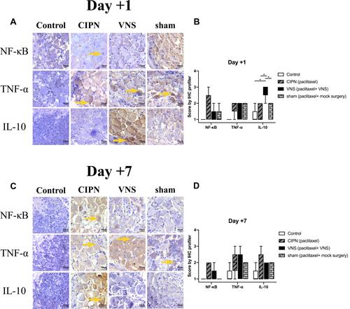
Figure 4 Pro- and anti-inflammatory regulators in dorsal root ganglia. Immunohistochemistry was performed on dorsal root ganglia tissue taken from CIPN rats treated with vagus nerve stimulation (VNS) or not. (A, C) Representative images of staining against NF-κB, TNF-α, and IL-10 on day +1 or +7. (B, D) Immunohistochemistry score was calculated using the IHC Profiler plugin in ImageJ (see Methods). Magnification, 20X. n = 5 per group per time point. Yellow arrows indicate areas of high expression. * p<0.05.