Figures & data
Figure 1 Experimental protocol. The evaluation was performed at four points: Baseline (pre-saline infusion; T0), Pre-intervention (stable painful state after saline infusion; T1), Post-intervention (3 minutes after intervention; T2) and 10 minutes after the pain had subsided (Recovery; T3). All subjects received hypertonic and isotonic infusions each day. The order of infusion type of saline (hypertonic/isotonic) and measurement (Experiment-A or -B) were randomized and balanced. Experiment-A contained 2 sessions which had 2 types of motor imagery interventions (ankle or finger) whereas Experiment-B had only 1 session (ankle motion imagery). Experiment-A measured H/M ratio, while Experiment-B recorded MVC force and RMS of the amplitude.
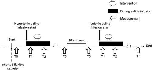
Figure 2 Experimental ankle pain model. (A) Set-up of the flexible catheter insertion. (B) Ultrasound image of the catheter inserted into Kager’s fat pad (A) Calcaneus, (B) Achilles tendon, Kager’s fat pad (yellow), catheter (white). *Tip of the catheter. (C) Syringe infusion pump attached to the catheter.
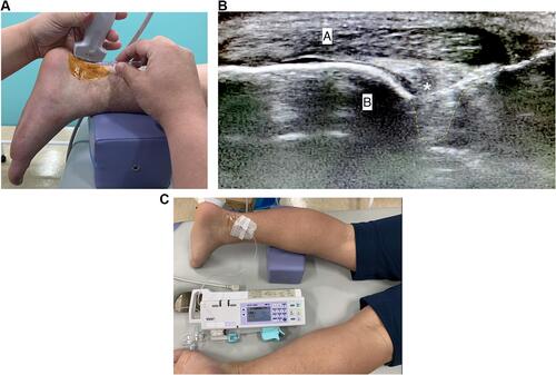
Table 1 Experimental Ankle Pain Intensity
Figure 3 Example of experimental protocol for a subject. The pain VAS after isotonic saline (blue line) and hypertonic saline (red line) infusions are indicated respectively. Time 0 sec shows the beginning of saline infusion.
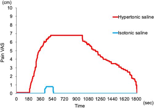
Table 2 H/M Ratio for Each Saline Infusion Over Time (Experiment-A)
Figure 4 Example of H-waves obtained from a subject in Experiment-A. Ankle movie was used for visually-assisted motor imagery. T0: Baseline, T1: Pre-intervention, T2: Post-intervention, T3: Recovery.
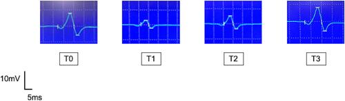
Figure 5 H/M ratio. Mean (SEM, n=10) H/M ratio after the infusion of hypertonic (open circle) or isotonic saline (black triangle). The data show H/M ratio after motor imagery of ankle motion (A) and hand motion (B). Although statistics were performed on raw values of the data, each number is indicated by the rate of change from Baseline (T0). Significantly different from T0 (*, NK: P<0.05), and T1 (†, NK: P<0.05). T0: Baseline, T1: Pre-intervention, T2: Post-intervention, T3: Recovery.
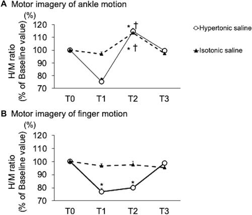
Figure 6 Maximal voluntary contraction force Mean (SEM, n=10) MVC after the infusion of hypertonic (open circle) or isotonic saline (black triangle). Although statistics were performed on raw values of the data, each number is indicated by the rate of change from Baseline (T0). Significantly different from T0 (*, NK: P<0.05), and T1 (†, NK: P<0.05). T0: Baseline, T1: Pre-intervention, T2: Post-intervention, T3: Recovery.
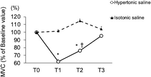
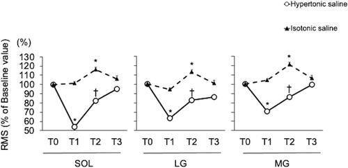
Figure 7 Contractile activity. Mean (SEM, n=10) RMS after the infusion of hypertonic (open circle) or isotonic saline (black triangle). Although statistics were performed on raw values of the data, each number is indicated by the rate of change from Baseline (T0). Significantly different from T0 (*, NK: P<0.05), and T1 (†, NK: P<0.05). T0: Baseline, T1: Pre-intervention, T2: Post-intervention, T3: Recovery.