Figures & data
Figure 1 The standing, trunk flexion and re-extension task.
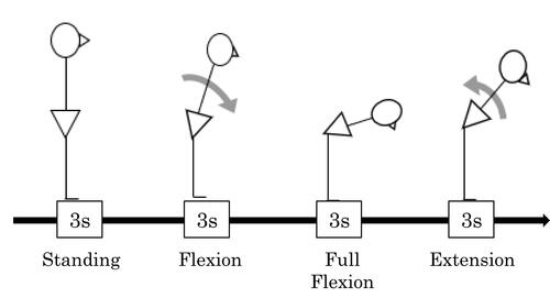
Figure 2 The approximate position of the EMG grid. The EMG electrode grid was placed 3 cm lateral to the lumbar spinous process on the bilateral erector spinae.
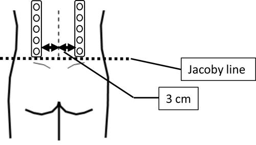
Table 1 Patient Characteristics
Table 2 Cross-Correlations at Zero Lag Indicating the Level of Cross-Sectional Associations Over Time Between Pain-Related Factors and Muscle Activity
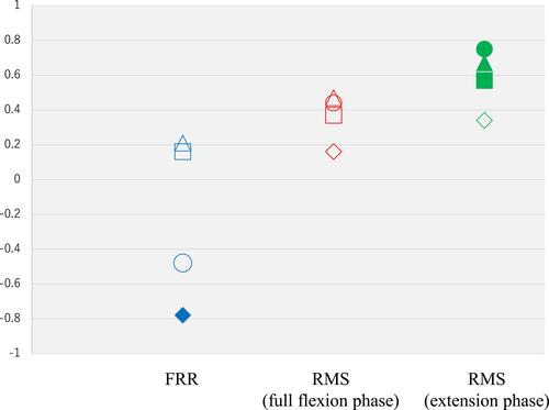
Figure 3 The correlation coefficients between each of the pain-related factors and muscle activity at zero lag.
Notes: Color-filled markers: indicate correlations that reached statistical significance. Unfilled markers: indicate correlations that not reached statistical significance. Circle markers: indicate correlations between the SFMPQ-2 and muscle activity. Triangle markers: indicate correlations between the PSFS and muscle activity. Square markers: indicate correlations between the OMSQ-12-psychological factor and muscle activity. Rhombus markers: indicate correlations between the FreBAQ and muscle activity. Abbreviations: SFMPQ-2, Short-Form McGill Pain Questionnaire-2; PSFS, Patient-Specific Functional Scale; FreBAQ, Fremantle Back Awareness Questionnaire; OMSQ-12, Örebro Musculoskeletal Screening Questionnaire-12; FRR, flexion relaxation ratio; RMS, root mean square.