Figures & data
Table 1 Primers for NGF
Figure 1 Estrogen downregulated rat chondrocytes NGF expression and release via ERα. (A) Rat primary chondrocytes were exposed to estrogen at a dose of 1 nM for 0, 12, 24, 48 or 72 hours. At 24 hours with 1 nM estrogen, expression of NGF mRNA was downregulated (0.7Ct, 0.55±0.13 fold) compared to the control. (B) Decrease in NGF protein levels were observed similarly to gene expression. (C) Rat primary chondrocytes were exposed to estrogen with or without ER inhibitors (MPP, PHTPP). Co-incubation with MPP, thereby preventing estrogen-ERα signaling could prevent estrogen-decreased NGF mRNA expression, while with PHTPP was not. (D) Co-incubation with MPP prevented estrogen-decreased NGF protein release. All data were shown as mean ± SEM from three independent experiments.
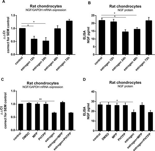
Figure 2 Estrogen decreased NGF expression and release in rat OA cartilage explants via ERα. (A) Rat cartilage explants were stimulated with estrogen (1nM) for 24 h, with or without ERα inhibitor (MPP, 20µM), after which mRNA was isolated to measure NGF expression. When exposed explants to estrogen, NGF mRNA expression was down-regulated about 0.8Ct (0.62±0.07 fold), similarly to chondrocytes response. Decreased reaction disappeared after estrogen combined with MPP. (B) NGF protein measured by ELISA matched mRNA changes. All data were shown as mean ± SEM from three independent experiments.
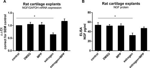
Figure 3 Estrogen down-regulate TGF-β1-stimulated and IL-1β-stimulated rat chondrocytes NGF mRNA expression and protein release. (A) Rat chondrocytes were stimulated for 24 hours with increasing concentrations of TGF-β1 (1, 10 and 20 ng/mL), and NGF mRNA expression was measured by quantitative qPCR. (B) NGF protein release was measured by ELISA. (C) Rat chondrocytes were stimulated for 24 hours with increasing concentrations of IL-1β (0.1, 1 and 10 ng/mL), and NGF mRNA expression was measured by quantitative qPCR. (D) NGF protein release was measured by ELISA. (E) TGF-β1 and IL-1β were added, respectively, into media with estrogen, with or without MPP. Estrogen almost suppressed TGF-β1 and IL-1β stimulated NGF mRNA expression. When cultured chondrocytes with estrogen, MPP, and TGF-β1/IL-1β, MPP resulted in estrogen-decreased phenomenon disappeared. (F) Co-incubated media were harvested and NGF protein was measured by ELISA. NGF protein levels paralleled with mRNA expression. All data were shown as mean ± SEM from three independent experiments.
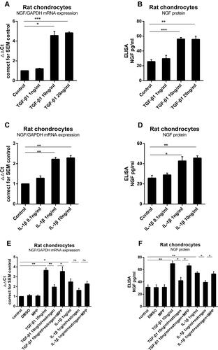
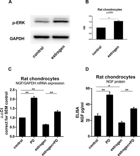
Figure 4 ERK1/2 was involved in the process of estrogen down-regulating NGF expression and release. (A) Protein was isolated from estrogen-treated chondrocytes, and p-ERK content was analyzed by WB, emerging a higher level than non-treated group. (B) WB results were quantified and presented in column graph style. Difference between two groups were seen clearly. (C) NGF mRNA expression in cultured with PD increased to 1.41Ct (2.07±0.1 fold) than non-treated. Estrogen-decreased NGF level was inhibited by PD. (D) NGF protein release paralleled with mRNA tendency. All data were shown as mean ± SEM from three independent experiments.