Figures & data
Figure 1 Unilateral laminotomy bilateral decompression via the posterior approach (schematic). The sequence of bony decompression was the ipsilateral lower lamina, upper spinous, contralateral lower lamina, ipsilateral lateral recess, lower spinous, and contralateral lateral recess.
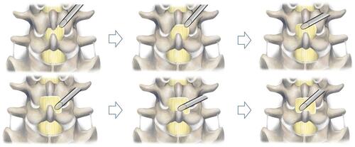
Figure 2 Sufficient neural decompression of full-endoscopic unilateral laminotomy bilateral decompression of L4/5. (A) Intraoperative endoscopic view of the dorsal side of neural decompression. (B) Intraoperative endoscopic view of the ventral side of neural decompression. (C) Postoperative CT (axial). (D) Postoperative CT (sagittal).
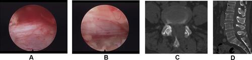
Table 1 Baseline Data in 2 Groups
Table 2 Surgical Outcomes in 2 Groups
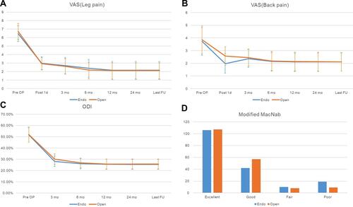
Figure 3 Clinical outcomes in the two groups. (A) VAS score (leg pain). (B) VAS score (back pain). (C) ODI was improved significantly in both groups. (D) Modified MacNab criteria of the two groups.