Figures & data
Figure 1 A panoramic radiograph taken at the time of the patient’s initial consultation revealed maxillary and mandibular partial edentulism. On the patient’s right side (left side of radiograph), there is an enlarged ankylotic mass fusing the mandible to the right base of the skull (circled in red), with impingement of the right maxillary tuberosity on the anterior aspect of the right mandible (yellow arrow). On the left mandible (right side of radiograph), there is a TMJ prosthetic implant with the mandibular condylar portion dislocated from the glenoid fossa component (blue arrow). Both mandibular coronoid processes are missing, having been surgically removed. The patient’s teeth are slightly apart – she can neither fully open nor close her mouth due to the bony fusion.
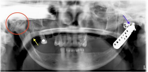
Figure 2 A post-operative panoramic after the patient underwent a series of three separate surgeries to correct her right mandibular bony ankylosis and previously placed failed left TMJ prosthesis. This radiograph depicts bilateral custom-made total TMJ prosthetic implants (TMJ Implants, Ventura, CA). The gaps between the prosthetic condyle and glenoid fossa plate (red arrows) represent the plastic insert on which the metallic condyle articulates. The embolization coil of the right maxillary artery is seen underlying the reconstructed TMJ (blue arrow). Temporary intermaxillary fixation wires are secured with screws (yellow arrows).
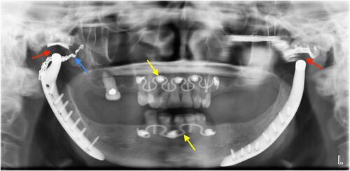
Figure 3 Timeline of events surrounding the manufacturing, clinical use, and ultimate recall of the VI PTIPI. Boxes above the timeline in from VI (red), scientific literature (green), and AAOMS actions (blue). Items below the timeline show FDA regulatory practices (yellow).
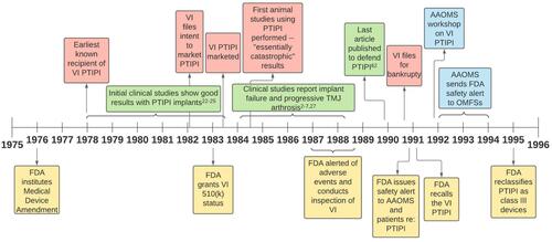
Figure 4 The rationale defending the patient’s allegations against Vitek, Inc. Proplast-Teflon interpositional implant (PTIPI) (red) and in defense of oral and maxillofacial surgeons (OMFS) (blue).
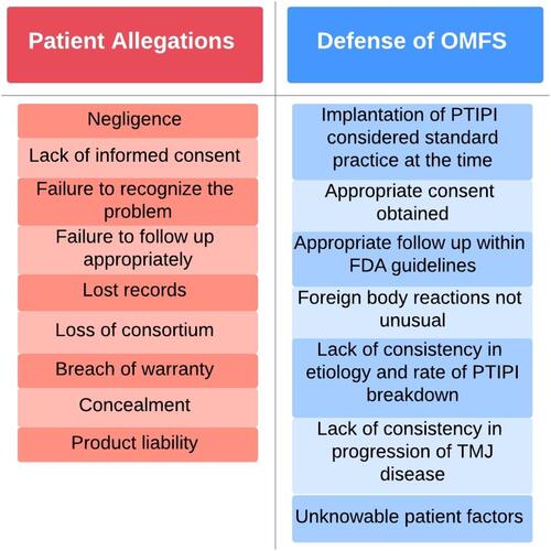
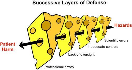
Figure 5 “Swiss cheese” model of failure, proposed and adapted by permission from BMJ Publishing Group Limited. Reason J. Human error: models and management. BMJ. 320, 768, copyright 2020.Citation80 In the case of the VI PTIPI, the risk associated with PTIPI managed to pass through “holes” despite layers of defense from the manufacturers, professional societies, hospitals, and the Food and Drug Association.