
Figures & data
Figure 1 Anatomical schematic diagram of the sensory pathway (left). After stroke, the body corresponds to the possible range of CPSP (right).
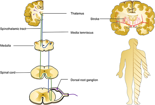
Table 1 Clinical Study of Spinal Thalamic Tract Injury in Central Pain After Stroke
Table 2 Molecular Mechanism Studies of CPSP