Figures & data
Figure 1 The endoscopic revision approach in patients of special herniations at L4/5.
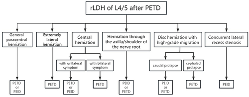
Table 1 Demographic Data of Patients
Table 2 Perioperative Indicators
Table 3 Preoperative, Follow-Up VAS, ODI, JOA Scores and Modified MacNab of Patients
Figure 2 Postoperative follow-up.
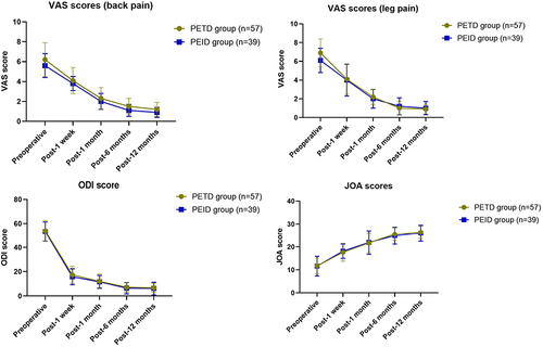
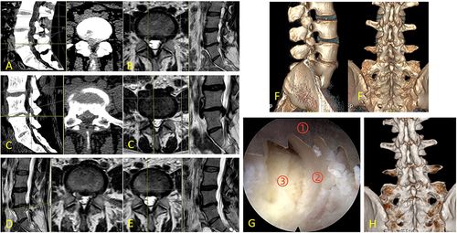
Figure 3 A 42-year-old female presented for the first time with “low back pain with pain in the left lower extremity”, and the lumbar MR showed a disc herniation at L45 (A). PETD was taken and the subsequent MR showed effective removal of the herniated disc (B) and (C) shows the removal of the articular process in the first operation. One year later, the patient presented with “low back pain with bilateral lower extremity numbness”, and the lumbar MR showed a large central herniation at L4/5 with cephalad prolapse (D). PETD was taken again, and (G)showed that the resection of the herniated disc reached all the way to the contralateral nerve root during the operation (① the contralateral nerve root, ② the ventral side of the spinal cord, ③ the residual disc). (H)is an intraoperative fluoroscopic image of the working channel, which shows that the grasping forcep has reached the position of the contralateral nerve root. (E and F) are lumbar MR at postoperative day 1 and 6 months, respectively.(I) shows that revision surgery removes the articular process to a greater extent, but still preserves part of the joint.
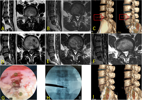
Figure 4 A 37-year-old woman’s first visit to the doctor was for “low back pain with numbness and pain in the right lower limb”, and the lumbar CT showed a disc herniation in the L4/5 (A), which was treated with PETD, and the follow-up MR showed that the herniated disc had been removed (B). The patient had a recurrence after 3 years, complaining of low back pain with bilateral lower limbs pain, and the lumbar CT and MR showed a disc herniation at L4/5 with lateral socket stenosis (C). PEID was used for revision, and (G) shows that part of the intraoperative lamina was resected with a visualized circular saw ((① the serration of circular saw, ② the vertebral plate, ③ the ligamentum flavum). (D and E) are postoperative MR on the first day and at 6 months, respectively, showing that the surgery was successful in removing the herniated disc. And (F–H) show the vertebral plates before and after revision.