Figures & data
Table 1 Demographic Characteristics of Enrolled Patients
Table 2 Comparison of Scores Between Two Groups
Figure 1 Postoperative scores of VAS-BP, VAS-LP and ODI significantly improved in both groups. p-value<0.001 in comparison between preoperative and all postoperative valued scores.
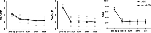
Figure 2 A 71-year-old man with ASD accepted PTED for L5-S1 disc herniation. (A) Preoperative anterior and lateral X-rays showing the L3-L5 having undergone PLIF surgery previously (B) Preoperative CT and MRI showing herniation of the intervertebral disc in the right of adjacent segment L5-S1 (yellow arrow) (C) Postoperative anterior and lateral X-rays showing the L5-S1 having undergone PTED (D) Postoperative CT and MRI showing the herniated intervertebral disc in the right side of L5-S1 having been removed (yellow arrow).
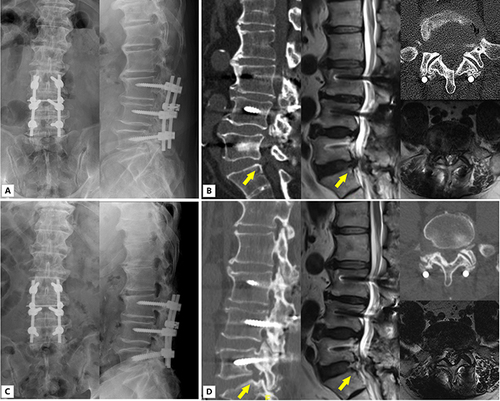
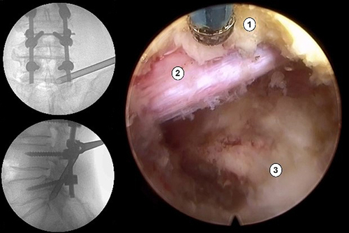
Figure 3 Intraoperative fluoroscopy shows the placement of the protective sleeve; Screenshot of Intraoperative endoscope shows the S1 nerve root after decompression. (1) ligamentum flavum; (2) S1 nerve; (3) intervertebral disc.