
Figures & data
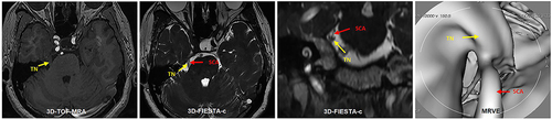
Table 1 MRI Findings and MVD Results of NVC
Figure 1 A 50-year-old female with left TN for 3 years. 3D-FIESTA-c axial (A) and 3D-TOF-MRA axial (B) showed vascular contact (red arrow) at the left trigeminal nerve (yellow arrow). MRVE image (C) showed the left Superior Cerebellar Artery (red arrow) compressing the trigeminal nerve from above (yellow arrow).
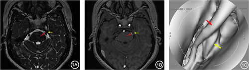
Figure 2 A 66-year-old female with right TN for over 5 years. 3D-FIESTA-c axial (A) and 3D-TOF-MRA axial (B) showed two branches of the right Superior Cerebellar Artery (red arrows) individually compressing the trigeminal nerve (yellow arrows). MRVE image (C) showed the two branches of the right Superior Cerebellar Artery (red arrows) separately compressing the trigeminal nerve from above (yellow arrows).
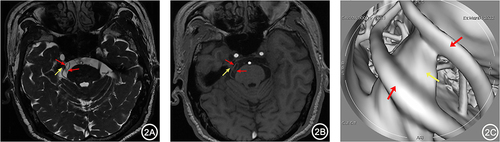
Figure 3 A 65-year-old male with right TN for 4 years. 3D-FIESTA-c axial (A) and coronal (B) showed close contact between the right Petrosal Vein (blue arrow) and the trigeminal nerve (yellow arrow) below. 3D-TOF-MRA axial (B) showed no apparent responsible vessels around the right trigeminal nerve (yellow arrow). MRVE image (D) showed the intimate relationship between the right Petrosal Vein (blue arrow) and the trigeminal nerve (yellow arrow).
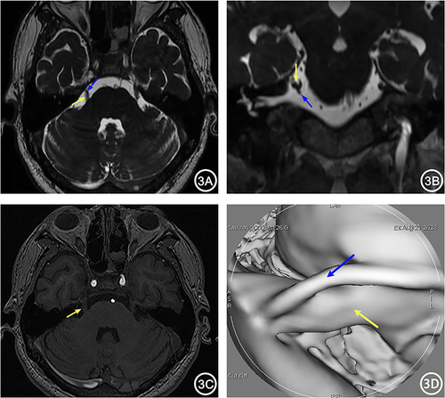
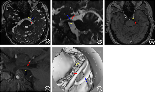
Figure 4 A 67-year-old female with left TN for 1 year. 3D-FIESTA-c axial (A) and sagittal (B) showed compression on the left trigeminal nerve (yellow arrow) by the Petrosal Vein (blue arrow) and Superior Cerebellar Artery (red arrow) from above. 3D-TOF-MRA axial (C) and sagittal (D) showed only the compression of the left trigeminal nerve by the Superior Cerebellar Artery (red arrow), with no other visible vascular. MRVE image (E) showed compression on the left trigeminal nerve by the Petrosal Vein (blue arrow) and Superior Cerebellar Artery (red arrow).
Data Sharing Statement
The data are available from the corresponding author on reasonable request.