
Figures & data
Figure 1 The course of the third occipital nerve as it traverses over the C2-3 facet joint.
Note: Used with permission from Mayo Foundation for Medical Education and Research © 2011.
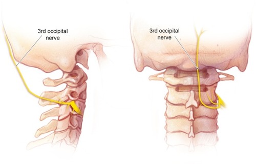
Figure 2 An anteroposterior radiograph of needle placement for third occipital nerve, C3-4, and C4-5 medial branch radiofrequency ablation.
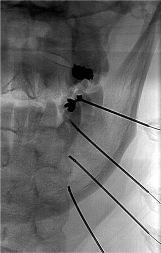
Figure 3 A lateral radiograph demonstrating the first needle placement for radio-frequency ablation of the third occipital nerve.
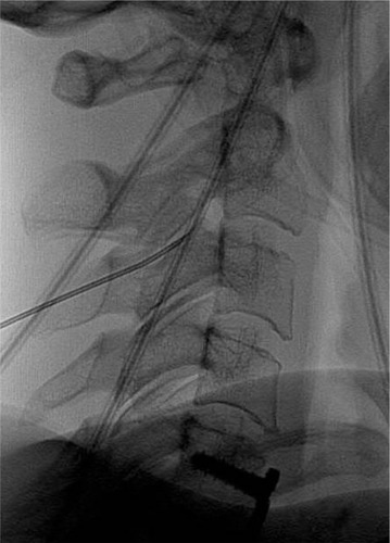
Figure 4 A lateral radiograph demonstrating the second (left) and third (right) needle placements for radiofrequency ablation of the third occipital nerve.

Table 1 Yearly numbers of patients with third occipital nerve pain and the number of patients requiring treatment