Figures & data
Figure 1 Fluoroscopic view of horizontal spread of contrast dye, prone position.
Note: The needle tip is seen near the inferior border of T12 rib on the left.
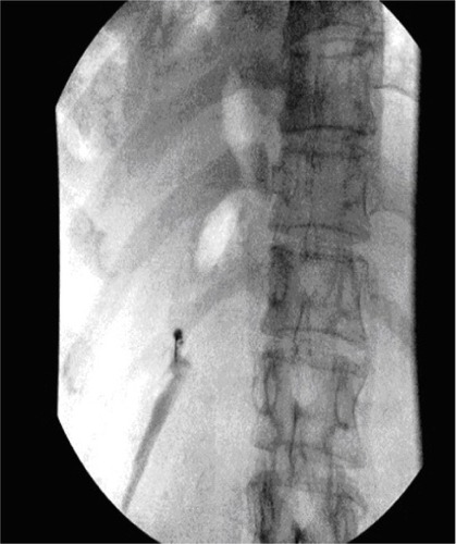
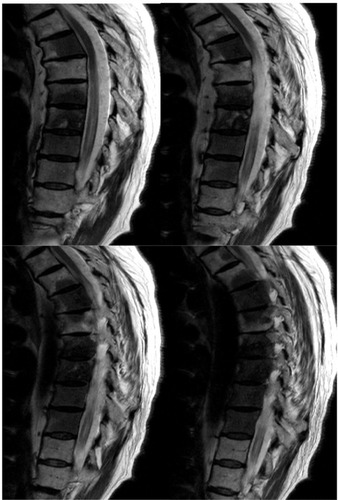
Figure 2 Magnetic resonance images of the thoracic spine, sagittal view, different sections, showing the thoracic body involvement.
Note: Extensive metastatic disease of the vertebral body can be seen.