Figures & data
Figure 1 Constitution of a nerve in transverse section.
Notes: Pictured is the fasciculate architecture that is created following the organization operated by the connective tissue that forms the sheaths of the nerve and the individual bundles of fibers. 1: Nerve bundles. 2: The endoneurium. 3: The perineurium. 4: Blood vessels. 5: Fat. 6: The epineurium. Reproduced with permission from Anastasi et al. AA VV, Anatomia dell’Uomo [Human Anatomy]. 4th ed. Milan: Edi. Ermes. Italian.Citation30 Copyright 2010 Edi. Ermes, Milano.
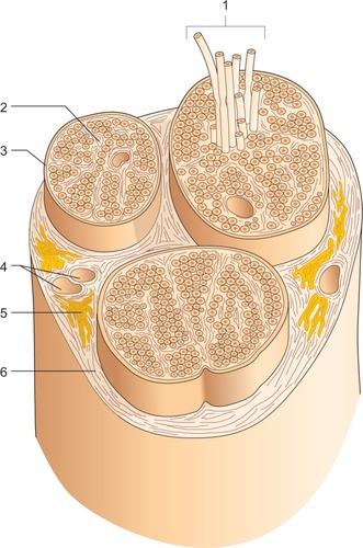
Figure 2 Location of the median and ulnar nerves in the region of the palm.
Notes: 1: The ulnar nerve; 2: the transverse carpal ligament; 3: deep palmar branch of the hand; 4: anastomosis between the median nerve and ulnar nerve; 5: digital nerves; 6: the median nerve. Reproduced with permission from Anastasi et al. AA VV, Anatomia dell’Uomo [Human Anatomy]. 4th ed. Milan: Edi. Ermes. Italian.Citation30 Copyright 2010 Edi. Ermes, Milano.
![Figure 2 Location of the median and ulnar nerves in the region of the palm.Notes: 1: The ulnar nerve; 2: the transverse carpal ligament; 3: deep palmar branch of the hand; 4: anastomosis between the median nerve and ulnar nerve; 5: digital nerves; 6: the median nerve. Reproduced with permission from Anastasi et al. AA VV, Anatomia dell’Uomo [Human Anatomy]. 4th ed. Milan: Edi. Ermes. Italian.Citation30 Copyright 2010 Edi. Ermes, Milano.](/cms/asset/5a532bd0-ccc1-4579-b434-4ce89c0e1162/djpr_a_89393_f0002_c.jpg)
Figure 3 Fascial treatment of the ulnar nerve in the peripheral nervous system.
Notes: A finger or fingers are placed on the nerve emergence at the elbow on the cubital tunnel, where it passes the ulnar nerve. The operator places his or her fingers on the fascial restriction identified previously by palpation, using different approaches, until the perceived resistance disappears or greatly decreases, inducing or stopping the preferential direction of the tissue.
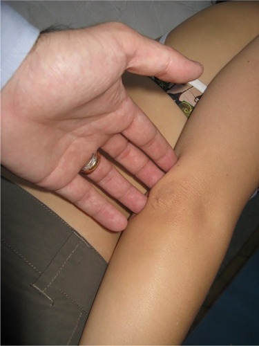
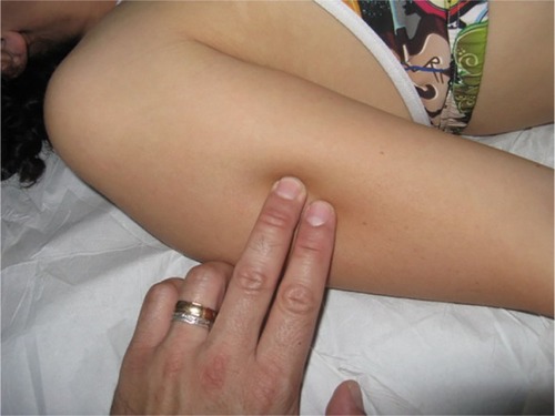
Figure 4 Fascial treatment of the radial nerve in the peripheral nervous system.
Notes: A finger or fingers are placed on the nerve emergence in the middle third lateral of the humerus, where the radial nerve passes. The operator places his or her fingers on the fascial restriction identified previously by palpation, using different approaches, until the perceived resistance disappears or greatly decreases, inducing or stopping the preferential direction of the tissue.