Figures & data
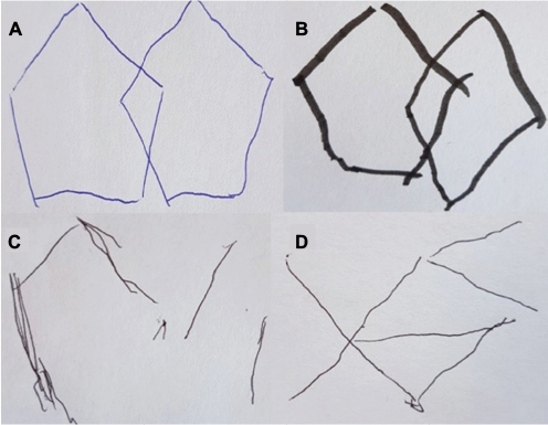
Figure 1 Visuospatial function is affected in Parkinson’s cognitive impairment.
Notes: Progressive difficulty copying interlocking pentagons. Based on level 1 MDS diagnostic criteria, patient “A” has normal cognition, patient “B” has PD-MCI and patients “C” and “D” have PDD.
Abbreviations: MDS, Movement Disorder Society; PD, Parkinson’s disease; PDD, PD dementia; PD-MCI, PD mild cognitive impairment.
Abbreviations: MDS, Movement Disorder Society; PD, Parkinson’s disease; PDD, PD dementia; PD-MCI, PD mild cognitive impairment.
Box 1 Summary of risk factors for PDD
Figure 2 The dopamine overdose hypothesis.
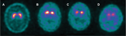
Notes: Progressive dorsal–ventral loss of striatal dopaminergic neurons is demonstrated on Dopamine transporter imaging using 123I-N-3-fluoropropyl-2beta-carbomethoxy-3beta-4-iodophenyl tropane single-photon emission computed tomography (123I-FP-CIT SPECT): substantia nigral projections to the dorsal putamen are lost early in PD (A), then the ventral putamen (B) and caudate in later stages (C and D). Dorsal putamen domains (executive function and working memory) are improved by dopaminergic stimulation early in the disease course, whereas the cognitive functions associated with the relatively preserved ventral striatum (reversal learning and motor sequence learning) are “overdosed” by dopaminergic stimulation at this stage.
Abbreviation: PD, Parkinson’s disease.
Abbreviation: PD, Parkinson’s disease.