Figures & data
Figure 1 Relationship between VSMC contraction and blood vessel diameter.
Notes: From a basal resting tone – and semi-contracted state – the VSMCs can either contract further, leading to a reduced blood vessel diameter and increased blood pressure, or relax, leading to a larger vessel diameter and reduced blood pressure. B, Basal resting tone, C, contraction, and R, relaxation of VSMCs.
Abbreviation: VSMC, vascular smooth muscle cell.
Abbreviation: VSMC, vascular smooth muscle cell.
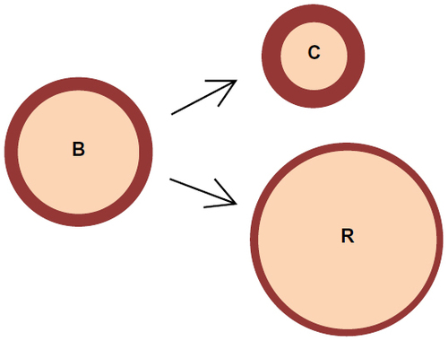
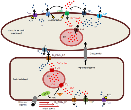
Figure 2 Modulation of membrane potential by K+ channels in vascular cells.
Notes: Endothelial cell: activation of KCa2.3/KCa3.1 channels in response to IP3R-mediated Ca2+ release from the ER after receptor stimulation or in response to Ca2+ influx through TRP channels, and communication to adjacent smooth muscle cell via gap junctions and KCa2.3/KCa3.1 signaling. Smooth muscle cell: activation of KATP, KV, VDCCs, and K2P channels. Activation of BKCa channels by RyR-mediated Ca2+ sparks from the SR leads to hyperpolarization of the membrane potential and closure of VDCCs.
Abbreviations: ATP, adenosine triphosphate; BKCa, large-conductance Ca2+-activated K+ channel; ER, endoplasmic reticulum; IP3, inositol 1,4,5-trisphosphate; IP3R, IP3 receptor; PLC, phospholipase C; PIP2, phosphatidyl-inositol-bisphosphate; K2P, two-pore domain K+ channel; KATP, ATP-sensitive K+; KCa, Ca2+-activated K+ channel; KV, voltage-gated K+ channel; RyR, ryanodine receptor; SR, sarcoplasmic reticulum; TRP, transient receptor potential; VDCC, voltage-dependent Ca2+ channel.
Abbreviations: ATP, adenosine triphosphate; BKCa, large-conductance Ca2+-activated K+ channel; ER, endoplasmic reticulum; IP3, inositol 1,4,5-trisphosphate; IP3R, IP3 receptor; PLC, phospholipase C; PIP2, phosphatidyl-inositol-bisphosphate; K2P, two-pore domain K+ channel; KATP, ATP-sensitive K+; KCa, Ca2+-activated K+ channel; KV, voltage-gated K+ channel; RyR, ryanodine receptor; SR, sarcoplasmic reticulum; TRP, transient receptor potential; VDCC, voltage-dependent Ca2+ channel.