
Figures & data
Figure 1 Representative histologic image from a needle biopsy of a 3.7 cm left lower lobe lung lesion (October 2015). H&E stained slides show well- to moderately differentiated adenocarcinoma with predominantly acinar growth pattern and associated psammomatous calcifications. (20x, H&E).
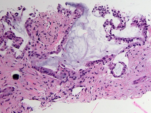
Figure 2 (A) ALK immunohistochemical staining of the needle biopsy specimen from October 2015, showing nearly diffuse expression of weak to moderate intensity. No background staining is present (10x, ALK, Novocastra 5A4). For comparison, a negative control is shown (B, 10x), consisting of the immunohistochemistry reaction without the primary ALK antibody; the lack of staining in the negative control (B) confirms that ALK staining in the patient sample (A) is due to the detection of the antigen. A positive control is also shown (C, 10x), showing diffuse ALK expression in a sample of an inflammatory myofibroblastic tumor, which is known to express the ALK protein.
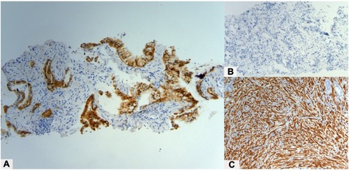
Figure 3 Integrative Genomics Viewer screenshot demonstrating next generation sequencing data (FoundationOne) from three specimens (TRF123157, specimen from October 2015; TRF166853, from June 2016; TRF232300, from May 2017). The specimens from June 2016 and May 2017 both show a CTNNB1 (NM_001904.4) c.133_134delTCinsGT, p.S45V dinucleotide substitution; however, this variant is not seen in the original specimen from October 2015.
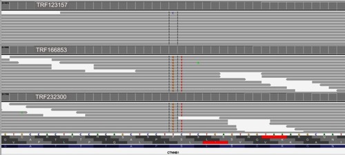
Figure 4 Timeline of clinical course, including comprehensive genomic profiling results and therapy.
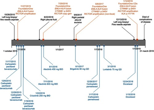
Table 1 Duration and best response of each treatment