Figures & data
Table 1 Relative yields, advantages, and shortcomings of currently available diagnostic techniques in the clincal workup of lung cancer
Figure 1 Virtual reconstruction of the endobronchial route to a lesion of interest showing an obvious bronchus sign leading to the tumor.
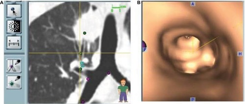
Figure 2 ENB procedure screen capture showing that an adenocarcinoma has been reached (green dot); (A–C) Sagittal, coronal, and axial views. The nodule is quite central and difficult to reach. (D) Indicates distance to the target and probe’s position. The patient required a preoperative diagnosis in order to be eligible for a clinical trial.
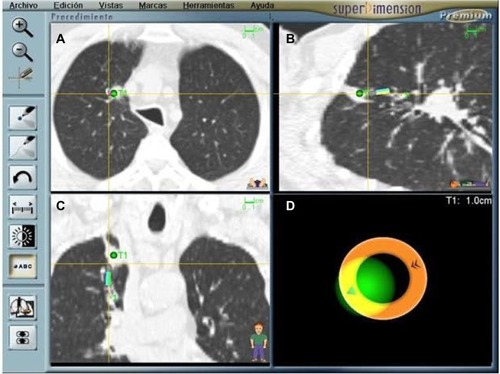
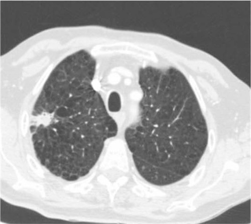
Figure 3 Spiculated nodule in a high-risk patient. ENB results were consistent with aspergilloma. Long-term follow-up has confirmed the benign diagnosis which spared this patient (with limited lung function) a risky surgical resection.