Figures & data
Figure 1 Triangle of Petit.
Figure 2 Suggested ultrasound probe positions for various approaches. (A) Initial scanning position for identification of linea alba and rectus abdominis, (B) probe position for subcostal approach, (C) probe position for lateral approach, (D) probe position for posterior approach.
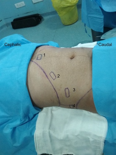
Figure 3 USG image at position 1 of .
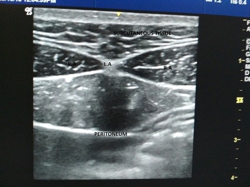
Figure 4 Subcostal TAP block. Arrow highlights the tap plane where the drug should be deposited.
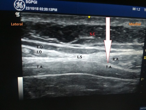
Figure 5 Subcostal TAP block: direction of needle and probe.
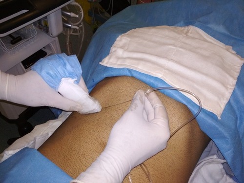
Figure 6 Lateral TAP block. Direction of needle and probe.
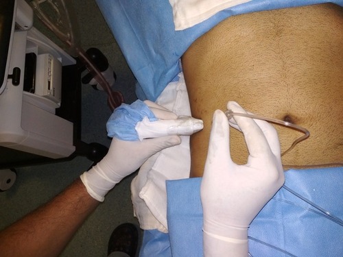
Figure 7 Lateral TAP block. Arrow highlights the tap plane where drug should be deposited.
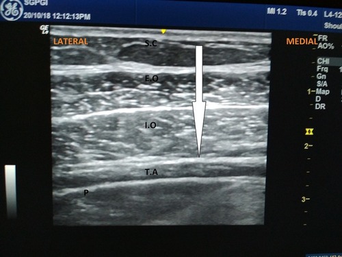
Figure 8 Posterior TAP block. Direction of needle and probe.
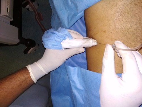
Figure 9 Posterior TAP block. Arrow highlights the TAP plane where drug should be deposited.
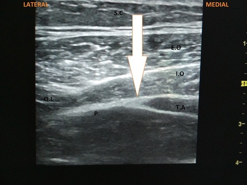
Table 1 Classification of USG guided TAP block
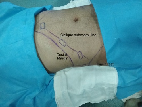
Figure 10 Direction of hydrodisscetion in oblique subcostal block. Initially subcostal TAP block is performed, then the needle moved along the oblique subcostal line.