Figures & data
Figure 1 Position of the ultrasound probe and the needle during the procedure of cervical plexus blocks.
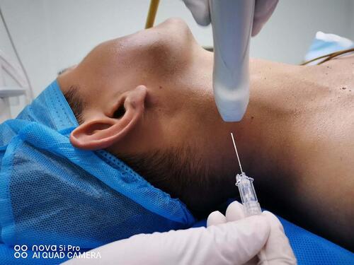
Figure 2 Ultrasound-guided deep and superficial cervical plexus block. (A) Ultrasound-guided transverse process block of the fourth cervical vertebrae. (B) Ultrasound-guided superficial cervical plexus block. Position of the needle and local anesthetic distribution was showed under the guidance of ultrasound during the procedure.
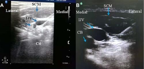
Table 1 Patient Demography and Comorbidities Data
Figure 3 Postoperative 48-hour pain trends (mean±SD). *P< 0.001, ANOVA with repeated measures. T4 indicates 10 min after extubation.
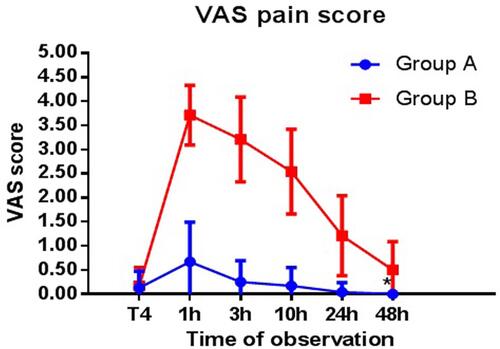
Table 2 Comparison of Anesthesia and Operation Time, and Drug Usage During the Perioperative Period
Table 3 Hemodynamics in the Operating Room (Invasive Systolic Arterial Blood Pressure [SABP], Diastolic Arterial Blood Pressure [DABP], and Heart Rate [HR])
Table 4 Statistics of Adverse Reactions
Table 5 Changes of Calcium, Phosphorus and Parathormone During the Perioperative Period
Figure 4 Calcium (A), phosphorus (B) and parathormone (C) values decreased after the surgery. *Indicates that there was no significant difference between the groups (P> 0.05).
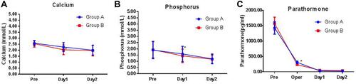
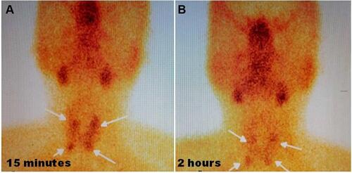
Figure 5 Localized nodules with increased uptake of radionuclides in the parathyroid region of bilateral thyroid lobes were considered as parathyroid hyperplasia or adenomas. 99mTc-MIBI was injected intravenously, and anterior imaging of the neck was performed 15 minutes after injection. Bilateral lobes of the thyroid gland were normal, and the radioactivity was evenly distributed. Radioactivity was aggregated in the parathyroid area of the upper and lower poles of the thyroid gland (A). After 2 hours delay, the distribution of thyroid radioactivity was lower than before, and there was still a slight localized radioactive accumulation in the parathyroid region of the upper and lower poles of the thyroid gland (B).