
Figures & data
Figure 1 Flowchart.
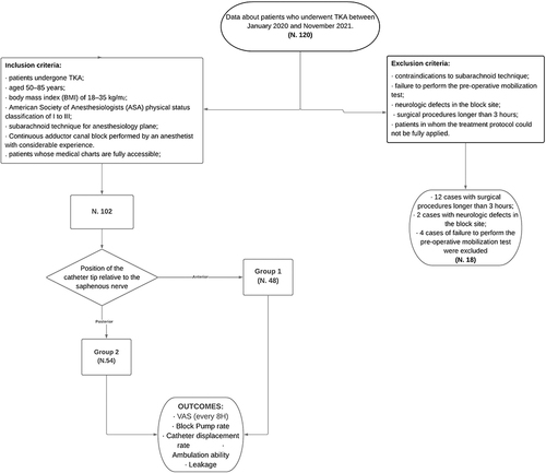
Figure 2 Continuous Adductor Canal Block (cACB): catheter tip anterior position. A 20 Gauge catheter was inserted through the split cannula anteriorly to the saphenous nerve. Sartorius muscle (S); saphenous nerve (N); femoral artery (A); local anesthetic (AL).
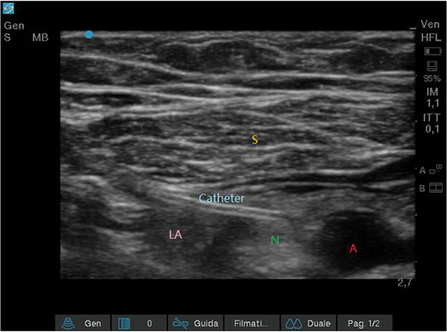
Figure 3 Continuous Adductor Canal Block (cACB): catheter tip posterior position. A 20 Gauge catheter was inserted through the split cannula posteriorly to the saphenous nerve. Sartorius muscle (S); saphenous nerve (N); femoral artery (A).
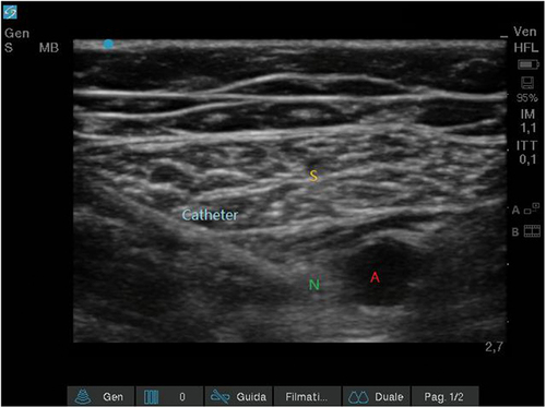
Table 1 Characteristics of Patients
Table 2 Outcomes
Table 3 VAS After Surgery