Figures & data
Table 1 CE-marked DCBs
Figure 1 Representative images of histological changes after IN.PACT™ Admiral™ DCB and Lutonix® DCB treatments, and histological scores of drug biological effects in the swine femoral artery model.
Abbreviations: DCB, drug-coated balloon; SMC, smooth muscle cell.
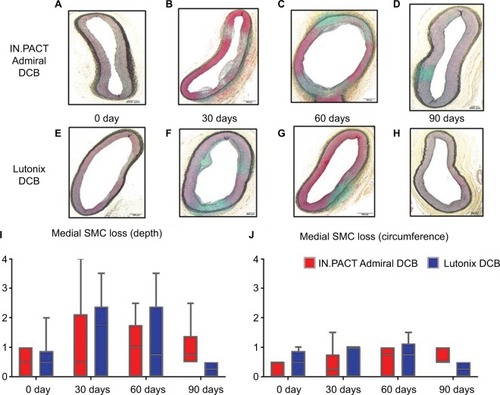
Table 2 Result from RCTs of currently available DCBsTable Footnotea
Figure 2 Representative images of downstream nontarget skeletal muscle and coronary band tissues from healthy swine after treatment of femoral arteries and downstream incidence of distal embolization after treatment with overlapping DCBs at 28 days follow-up.
Source: Reproduced with permission from Reprinted from Journal of Vascular and Interventional Radiology, 27, Frank D. Kolodgie, Erica Pacheco, Kazuyuki Yahagi, Hiroyoshi Mori, Elena Ladich, Renu Virmani, Comparison of Particulate Embolization after Femoral Artery Treatment with IN.PACT Admiral versus Lutonix 035 Paclitaxel-Coated Balloons in Healthy Swine, 1676-1685.e2., © 2016, with permission from Elsevier. And Reprinted from Journal of Vascular and Interventional Radiology, Sho Torii, Hiroyuki Jinnouchi, Atsushi Sakamoto, Maria E. Romero, Frank D. Kolodgie, Renu Virmani, Aloke V. Finn, Comparison of Biologic Effect and Particulate Embolization after Femoral Artery Treatment with Three Drug-Coated Balloons in Healthy Swine Model, Epub ahead of print., © 2018, with permission from Elsevier.Citation68
Abbreviation: DCBs, drug-coated balloons.
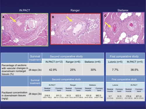
Figure 3 Primary patency and LLL at 12 months after the treatment in eight groups that were classified according to circumferential and longitudinal distribution of calcification.
Source: Reprinted by permission from Springer Nature Customer Service Centre GmbH: Springer Nature CardioVascular and Interventional Radiology Fanelli F, Cannavale A, Gazzetti M, et al. Calcium burden assessment and impact on drug-eluting balloons in peripheral arterial disease. Cardiovasc Intervent Radiol. 2014;37(4):898–907, © 2014.Citation59
Abbreviation: LLL, late lumen loss.
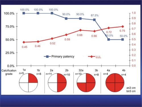
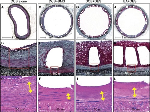
Figure 4 Representative histologic images of the biologic responses to DCB + DES, BA + DES, DCB + BMS, and DCB alone in swine iliofemoral arteries.
Source: Reprinted from Journal of Vascular and Interventional Radiology, 29, Sho Torii,Kazuyuki Yahagi, Hiroyoshi Mori, Emanuel Harari, Maria E. Romero, Frank D. Kolodgie, Brandt Young, Anthony Ragheb, Renu Virmani, Aloke V. Finn, Biologic drug effect and particulate embolization of drug-eluting stents versus drug-coated balloons in healthy swine femoropopliteal arteries, 1041–1049., © 2018, with permission from Elsevier.Citation61
Abbreviations: BA, balloon angioplasty; BMS, bare metal stents; DCB, drug-coated balloon; DES, drug-eluting stents; PTA, percutaneous transluminal angioplasty; SMC, smooth muscle cell.