Figures & data
Table 1 Electrodiagnostic findings of patients
Table 2 Severity-grading classification of ulnar neuropathy based on electrodiagnostic findings
Figure 1 Position of elbow in ultrasonography (from left to right) at level of medial epicondyle, 2 cm above it, and 2 cm below it, respectively.
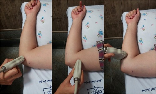
Figure 2 Ulnar nerve cross-section area on ultrasonography.
Note: (A) At level of medial epicondyle (arrow, ulnar nerve; arrowhead, medial epicondyle; star, olecranon); (B) 2 cm above medial epicondyle (arrow, ulnar nerve; star, medial epicondyle); (C) 2 cm below medial epicondyle (arrow, ulnar nerve; stars, two heads of flexor carpi ulnaris muscle).
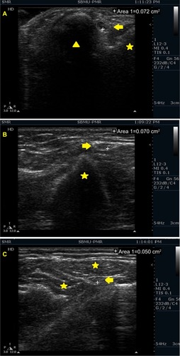
Table 3 Demographic characteristics of control and patient groups
Table 4 Distribution of ulnar nerve sonographic parameters between control and patient groups
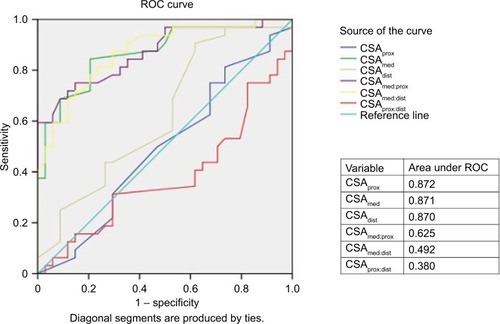
Figure 3 Receiver-operating characteristic (ROC) curve for diagnostic value of ultrasonographic parameters in ulnar neuropathy.
Abbreviations: CSA, cross-sectional area; dist, distal to medial epicondyle; med, at the level of medial epicondyle; prox, proximal to medial epicondyle.