
Figures & data
Figure 1 Barrier-forming properties of SSPCF: TEER and Lucifer yellow assays. (A) SSPCF treatment caused a significant decrease in resistance after 1 day and a significant increase in resistance after 3 days, as compared to saline treatment. The increase in resistance at day 3 indicates maintenance of epithelium integrity. (B) As compared to saline treatment, SSPCF treatment did not cause any significant change in the membrane permeability measured by the LY permeability assay. This further indicates the maintenance of the integrity and functionality of the epithelium. **p<0.01. Error bars represent SEM.
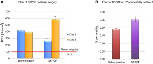
Figure 2 Secreted lactate dehydrogenase profile after treatment with SSPCF. Levels of LDH were measured in untreated, SSPCF-treated and Triton X-100-treated MucilAir™ epithelial cells for 1–4 days. SSPCF treatment did not cause an increase in the secreted LDH levels as compared to untreated tissues. This indicates the safety of SSPCF on the nasal epithelium in terms of cytotoxicity. The values were normalized to the levels of secreted LDH in the Triton-X 100-treated tissues (100%).
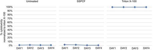
Figure 3 Effect of SSPCF on IL-8 secretion. Levels of IL-8 were measured in an ELISA assay in untreated, SSPCF-treated and Cytomix-treated MucilAir™ epithelial cells. SSPCF treatment did not cause a significant increase in the secreted IL-8 levels as compared to untreated tissues indicating the safety of use of SSPCF on the nasal epithelium in terms of pro-inflammation. Error bars represent SEM.
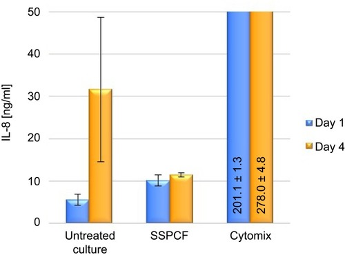
Figure 4 Mucociliary clearance after treatment with SSPCF. Mucociliary clearance rates in untreated, SSPCF-treated, and isoproterenol-treated tissues. Both on Day 1 and Day 4, SSPCF and isoproterenol treatments increased MCC rates, as compared to untreated cultures. The differences between SSPCF and isoproterenol on days 1 and 4 did not reach the statistical significance (Day 1: p=0.538; Day 4: p=0.717). This indicates an improvement in the mucociliary clearance upon SSPCF treatment. *p<0.05, **p<0.01, ***p<0.001. Error bars represent SEM.
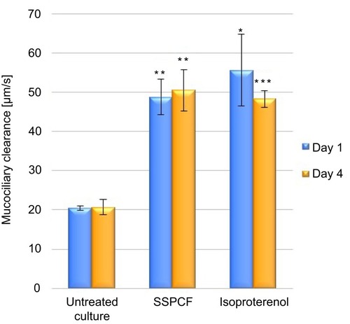
Figure 5 Evaluation of the ATP release and cell morphology effects of SSPCF. (A) ATP release (nM) after 2 mins of treatment following 5 mins of hypotonic saline solution pre-application. Hypotonic solution + saline treatment caused a significant increase in ATP release, as compared to saline-only control. On the other hand, the amount of ATP released upon hypotonic solution + SSPCF treatment was significantly lower compared to hypotonic solution + saline treatment. ***p<0.001. (B) Immunohistochemical staining of the tissues 2 mins after treatment, by AQP3 antibody and Alcian blue. These data indicate that SSPCF treatment helps recover the tissues from hypotonic stress. Error bars represent SEM.
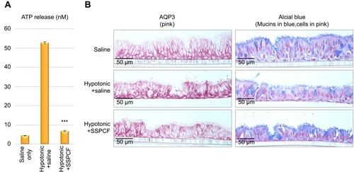
Table S1 TEER
Table S2 LY
Table S3 LDH
Table S4 IL-8
Table S5 MCC
Table S6 ATP