Figures & data
Figure 1 Distribution in brain tissue and blood concentration of coumarin after intravenous or intranasal administration of coumarin-loaded MPEG-PCL micelles.
Notes: (A) Coumarin levels in the brain tissue of tumor-inoculated rats. Rats were sacrificed at 1 hour (following intranasal administration) or 24 hours (following intravenous administration) after the injection of coumarin-loaded MPEG-PCL micelles (dose corresponding to 20 μg of coumarin). Each bar represents the mean ± SE (n=3). **P,0.01. (B) Coumarin concentration in blood after administration of coumarin solution or coumarin-loaded MPEG-PCL (dose corresponding to 20 μg of coumarin). Each point represents the mean ± SE (n=3). **P,0.01 versus intranasally administered MPEG-PCL.
Abbreviations: MPEG-PCL, methoxypolyethylene glycol-polycaprolactone; SE, standard error; h, hours; PEG, polyethylene glycol.
Abbreviations: MPEG-PCL, methoxypolyethylene glycol-polycaprolactone; SE, standard error; h, hours; PEG, polyethylene glycol.
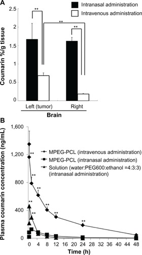
Figure 2 Structure of Tat-modified MPEG-PCL micelle.
Note: Carboxylic group in cell-penetrating peptide Tat was conjugated to the hydroxyl group in MPEG-PCL by esterification.
Abbreviations: MPEG, methoxypolyethylene glycol; PCL, polycaprolactone; CPP, cell-penetrating peptide; G, glycine; R, arginine; K, lysine; Q, glutamine.
Abbreviations: MPEG, methoxypolyethylene glycol; PCL, polycaprolactone; CPP, cell-penetrating peptide; G, glycine; R, arginine; K, lysine; Q, glutamine.
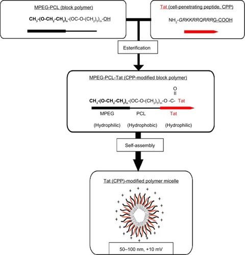
Figure 3 Biodistribution of coumarin in rats after intranasal administration of coumarin-loaded MPEG-PCL-Tat micelles.
Notes: Rats were sacrificed 1 hour (Image) or 4 hours (Image) after injection of coumarin-loaded MPEG-PCL-Tat micelles, or 1 hour (Image) following the injection of coumarin solution (dose corresponding to 20 μg of coumarin). Each bar represents the mean ± SE (n=3). nsP.0.05, **P,0.01, *P,0.05.
Abbreviations: MPEG-PCL, methoxypolyethylene glycol-polycaprolactone; SE, standard error; ns, not significant.
Abbreviations: MPEG-PCL, methoxypolyethylene glycol-polycaprolactone; SE, standard error; ns, not significant.
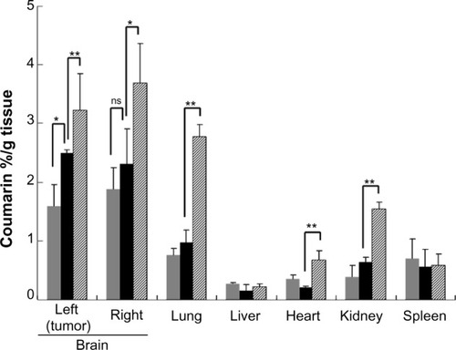
Figure 4 Distribution of FAM-siRNA in slices of nasal mucosa after intranasal administration of naked FAM-siRNA and MPEG-PCL-Tat/FAM-siRNA.
Notes: Rats were sacrificed 15 minutes after intranasal administration of naked FAM-siRNA (A) or MPEG-PCL-Tat/FAM-siRNA (B) (dose corresponding to 40 μg of FAM-siRNA, N/P=15). Untreated rats (C) were sacrificed at the same time point. Olfactory mucosa tissue was enucleated. Blue fluorescence represents the nuclei stained by Hoechst, and green fluorescence represents FAM-siRNA. Scale bar =200 μm. 6-Carboxyfluorescein-aminohexyl (FAM)-siRNA (Cosmo Bio Co., Ltd., Tokyo, Japan) as a fluorescent-labeled siRNA.
Abbreviations: OE, olfactory mucosa epithelium; LP, lamina propria; NC, nasal cavity; MPEG-PCL, methoxypolyethylene glycol-polycaprolactone; N/P, ratio of amine to nucleic acid; FAM, 6-Carboxyfluorescein-aminohexyl; siRNA, small interfering ribonucleic acid.
Abbreviations: OE, olfactory mucosa epithelium; LP, lamina propria; NC, nasal cavity; MPEG-PCL, methoxypolyethylene glycol-polycaprolactone; N/P, ratio of amine to nucleic acid; FAM, 6-Carboxyfluorescein-aminohexyl; siRNA, small interfering ribonucleic acid.
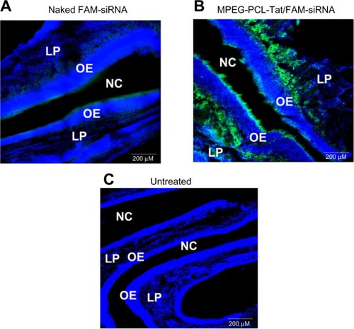
Figure 5 siRNA distribution in the olfactory bulb tissue after intranasal administration.
Notes: Rats were sacrificed 15 minutes after intranasal administration of naked FAM-siRNA (A) or MPEG-PCL-Tat/FAM-siRNA (B) (dose corresponding to 40 μg of FAM-siRNA, N/P=15). Each olfactory bulb was enucleated. Blue fluorescence represents the nuclei stained by Hoechst, and green fluorescence represents FAM-siRNA. Scale bar =200 μm. 6-Carboxyfluorescein-aminohexyl (FAM)-siRNA (Cosmo Bio Co., Ltd., Tokyo, Japan) as a fluorescent-labeled siRNA.
Abbreviations: GL, glomerular layer; EPL, external plexiform layer; MCL, mitral cell layer; GCL, granule cell layer; siRNA; small interfering ribonucleic acid; MPEG-PCL, methoxypolyethylene glycol-polycaprolactone; N/P, ratio of amine to nucleic acid; FAM, 6-Carboxyfluorescein-aminohexyl (FAM)-siRNA (Cosmo Bio Co., Ltd., Tokyo, Japan) as a fluorescent-labeled siRNA..
Abbreviations: GL, glomerular layer; EPL, external plexiform layer; MCL, mitral cell layer; GCL, granule cell layer; siRNA; small interfering ribonucleic acid; MPEG-PCL, methoxypolyethylene glycol-polycaprolactone; N/P, ratio of amine to nucleic acid; FAM, 6-Carboxyfluorescein-aminohexyl (FAM)-siRNA (Cosmo Bio Co., Ltd., Tokyo, Japan) as a fluorescent-labeled siRNA..
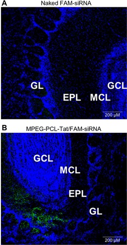
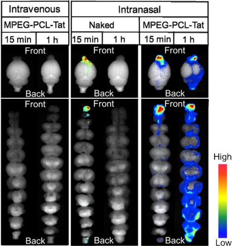
Figure 6 Dynamics of MPEG-PCL-Tat complex in brain tissue following intranasal or intravenous administration.
Notes: Rats were sacrificed and brains were enucleated at each point after intranasal or intravenous administration of naked Alexa-dextran or Alexa-dextran/MPEG-PCL-Tat (dose corresponding to 40 μg of Alexa-dextran). Each sample was observed using Maestro™ (Kurabo, Industries, Ltd., Osaka, Japan). Anionic dextran labeled with Alexa Fluor® 678 (molecular weight: 10,000), (Alexa-dextran; Life Technologies, Carlsbad, MA, USA).
Abbreviations: MPEG-PCL, methoxypolyethylene glycol-polycaprolactone; h, hour; min, minutes.
Abbreviations: MPEG-PCL, methoxypolyethylene glycol-polycaprolactone; h, hour; min, minutes.