Figures & data
Figure 1 The vestibular system.
Notes: Used with permission of Mayo Foundation for Medical Education and Research. All rights reserved. The vestibular system is composed of the three semicircular canals, the utricle, and the saccule (not labeled but adjacent to the utricle), which are filled with endolymph fluid. Hair cells located in the cupula have stereocilia that detect endolymph flow in response to angular or linear acceleration.
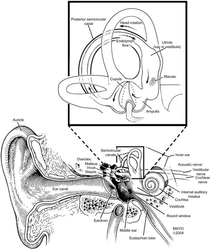
Figure 2 Motion detection in the vestibular system.
Notes: Adapted from Current Biology; 21(19); Straumann D, Bockisch C; Neurophysiology: vertigo in MRI machines; R806–R807; Copyright © 2011, with permission from Elsevier.Citation9 As the head moves in space, the endolymph flows in an opposite direction within the semicircular canal. This flow creates hydrodynamic pressure, which is detected by hair cells in the cupula.
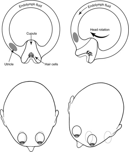
Figure 3 The Lorentz force.
Notes: Adapted from Current Biology; 21(19); Straumann D, Bockisch C; Neurophysiology: vertigo in MRI machines; R806–R807; Copyright © 2011, with permission from Elsevier.Citation9 The Lorentz force arises in response to current flow that is induced by the magnetic field. Depending on the orientation of the subject’s head, the Lorentz force can cause deflection of the hair cells in the cupula, which can cause a sense of movement, when in fact the subject is stationary. This is experienced as vertigo (illustration adapted from Straumann and Bockisch,Citation9 with permission of the author and publisher).
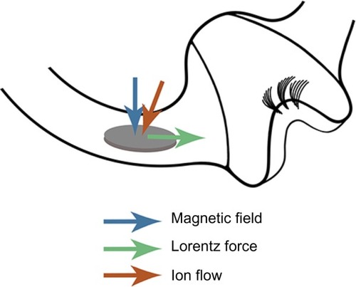
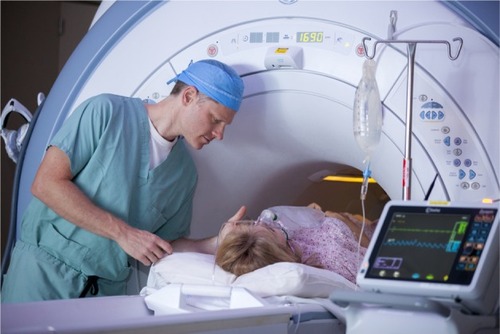
Figure 4 An anesthesia provider leans into the MRI bore while attending to a patient, exposing him to a much stronger magnetic field and increasing his risk of vertigo (photo by Peter Pallagi).