Figures & data
Figure 1 Schematic diagram of biosensor with target analyte (A) and nontarget analyte (B).
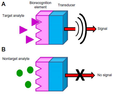
Figure 2 Structure of “nanobiosensor for virus detection” segmentation.
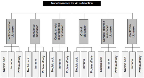
Figure 3 Strategies for electrochemical biosensing of viral pathogens consist of four steps.
Notes: Four strategies for electrochemical biosensing of viral pathogens are as follows: (A) target parts of virion, (B) modification of electrode (sensor) by biorecognition element, (C) isolation of targets, (D) signal detection or detection of signal after amplification.
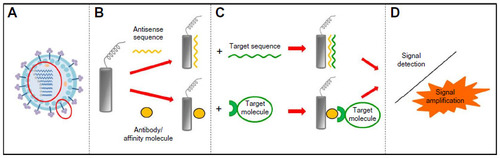
Figure 4 Impedance biosensor for measurement of immunoreaction coupled with red blood cell (RBC) amplification.
Notes: The protocol consisted of three parts: (A) gold electrode surface modification by polyclonal anti-N1 antibody, (B) H5N1virions binding and detection, and (C) RBC amplification. RBCs were used as biolabels to attach to captured H5N1 to amplify impedance signal.
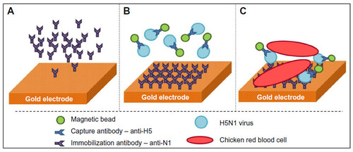
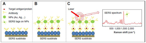
Figure 5 Surface-enhanced Raman scattering (SERS) biosensor for antigen/specific protein detection.
Notes: (A) SERS substrate modification by antitarget antibody, (B) target isolation, followed by binding of nanoparticles (NPs), lebeled by SERS tag, and SERS-tag detection (C).