Figures & data
Figure 1 (A) First EEG pattern of the patient: atypical delta wave complexes on our patient’s EEG. Left postero-temporal focal lesion with polymorph delta waves (1 – shaded). (B) Second EEG pattern of the patient: alpha-basic rhythm with 8–9 cycles per second and with diffuse irritative changes, unmodified during eye opening, with asymmetric slow sharp wave discharges in the C-T region bilaterally (1 and 2 – shaded). (C) Normal EEG pattern for comparison: a basic activity with subalpha-theta waves, without pathological changes and asymmetry. (A and B) Registrations with time constant 0.10 s, high frequency filter 30.0 Hz, notch filter: yes, sensitivity: 5.0 µV/mm. (C) Registration with time constant 0.10 s, high pass filter 5.30 Hz, low pass filter 20.0 Hz, notch filter: yes.
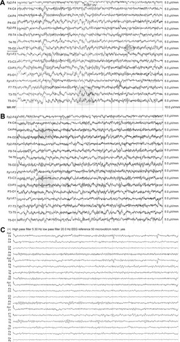
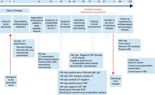
Figure 2 Timeline graph of the main events in our patient’s disease course.