Figures & data
Figure 1 Duplex ultrasound (B-mode) shows narrowing of left intervertebral VA. The asterisk indicates the false lumen caused by intramural hematoma.
Abbreviation: VA, vertebral artery.
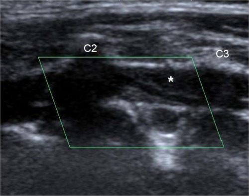
Figure 2 Color flow longitudinal duplex ultrasound highlights the severe luminal narrowing of left intervertebral VA due to intramural hematoma.
Abbreviation: VA, vertebral artery.
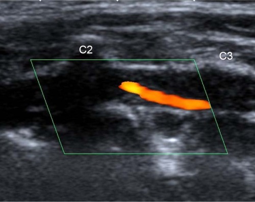
Figure 3 The coronal fat-saturated T1-weighted MRI sequence shows dissection of the left VA (arrow heads).
Abbreviations: MRI, magnetic resonance imaging; VA, vertebral artery.
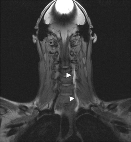
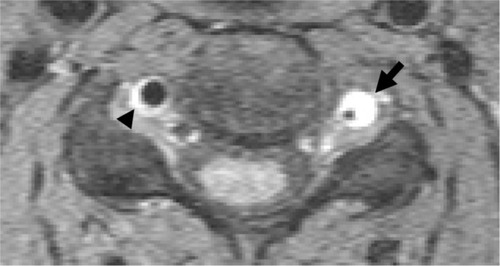
Figure 4 The axial fat-saturated T1 MRI sequence shows dissection of the left (arrow) and dissection of the right VA (arrow head).
Abbreviations: MRI, magnetic resonance imaging; VA, vertebral artery.