Figures & data
Figure 1 Diagram showing some of the possible clinical evolution that can be interpreted as natural history of TS or outcome from non-surgical therapeutic interventions based on YGTSS as a severity measure. This diagram intends to illustrate the current indications for DBS according to earlier and latest criteria. (A) Clinical resolution of TS symptoms. (B) Presence of tics that do not resolve spontaneously or are kept stable under non-surgical treatments. (C) Classical indication for DBS based on the severity of disease and age (18 years). (D) Latest proposed indication for DBS based on severity as a determinant factor even before 18 years of age.
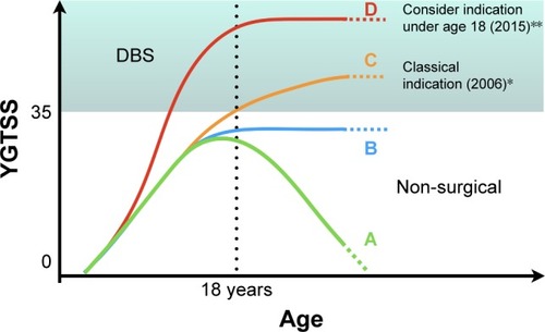
Table 1 Clinical criteria for the indication of DBS in Tourette’s syndrome
Table 2 Summary of the studies: level III evidence
Table 3 Summary of studies: level IV evidence
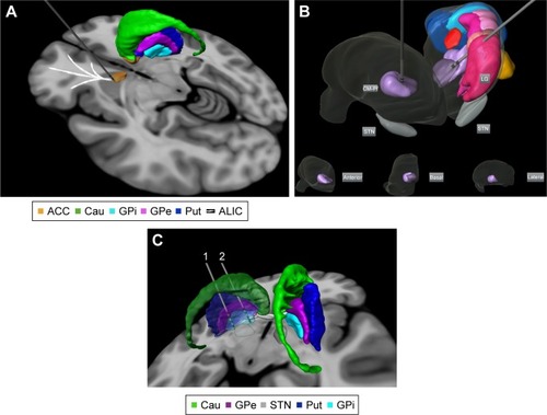
Figure 2 Targets proposed for DBS treatment in Tourette’s syndrome: (A) anterior limb of internal capsule/accumbens; (B) bilateral centromedian-parafascicular complex targeted in an anterolateral thalamic view (basal, anterior, and lateral thalamic views of the right thalamus are displayed for localization within the thalamus); and (C) different parts of GPi. Electrode 1 is located in the posteroventrolateral GPi and electrode 2 is located in the anteromedial GPi. The 3D representations are histological postmortem reconstructions of the nuclei from the University of São Paulo – Würzburg Atlas of the Human Brain (Alho et al, 2018Citation96).